2.2 POJA-L987
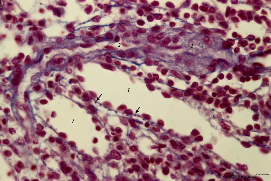
Title: Red pulp of spleen with perfused venous sinusoids (human)
Description: Stain: Azan.
Cross-sectioned venous sinuses with splenic cord (1). The wall of a sinusoid is composed of elongated rod-like endothelial cells that are orientated parallel to each other in the long axis of the sinus. There is a discontinuous pale-stained basement membrane (difficult to observe in light microscopy). Reticular fibres are deep-blue stained and are distributed everywhere. They are spirally wrapped fibres reinforcing the sinusoids.
At (→) the blue-stained dots of cross sectioned reticular fibres appear clearly surrounding a venous sinus.
(2): The wall of the arterioles contains myocytes.
Keywords/Mesh: lymphatic tissue, spleen, sinus, red pulp, histology, POJA collection

