
5.4.2 POJA-La0083+L2493+
4272+2491.
Macula densa cells of the juxtaglomerular apparatus in the kidney VIII
5.4.2 POJA-La0083+L2493+4272+2491
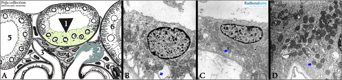
Title: Macula densa cells of the juxtaglomerular apparatus in the kidney VIII
Description:
(A): Glomerulus, electron microscopy scheme, human.
(1) Macula densa.
(2) JG cells in the wall of the afferent arteriole.
(3) Efferent arteriole.
(4) Goormaghtigh cells or lacis cells or Polkissen cells are extraglomerular mesangial cells.
The lacis cells are part of the juxtaglomerular apparatus that also comprises the macula densa (1) in the cortical thick
ascending limb of the distal tubule and the JG cells (2).
The JG apparatus controls the blood pressure through the Renin-Angiotensin-Aldosterone system.
(5) Distal convoluted tubule (pars contorta II).
(6) Proximal convoluted tubule (pars contorta I).
(B - D): Macula densa, electron microscopy, human. The macula densa cells are attached on the basal lamina of the tubule
with long and slender foot processes and are rich in mitochondria.
The degree of basal interdigitations (basal labyrinth, * * *) as well as the distinct presence of Golgi areas subnuclearly is related
to the activity of the macula densa cells.
Keywords/Mesh: urinary system, kidney, glomerulus, JG apparatus, macula densa, Goormaghtigh cell, lacis cell, histology, electron microscopy, POJA collection