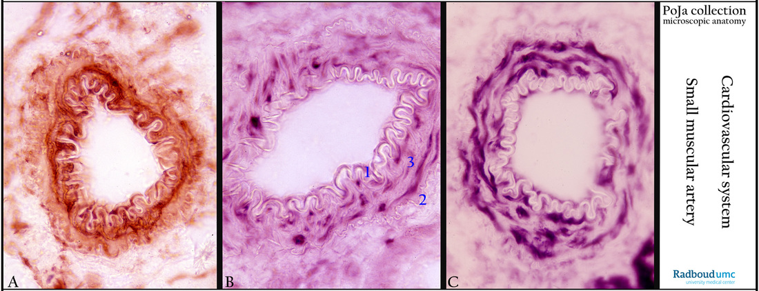
13.1 POJA-L4633+4634+4574
Title: Enzyme histochemistry of smooth muscle cells of blood vessels (mouse).
Description:
(A): ATPase staining (liver) of the muscular cells in the wall of the muscular artery. Note the folded white membrane elastica interna. VSM (=vascular smooth muscle) is primarily an oxidative tissue, more than 70 % of its ATP is obtained by oxidative phosphorylation. Compared to skeletal and cardiac muscles smooth muscle need less ATP to work efficiently.
(B): GPDH staining (kidney) of the muscle cells (3) in the wall of the muscular artery. (1) Membrana elastica interna. (2) Membrane elastica externa. The cytosolic glucose-6-phosphate dehydrogenase (G6PDH) is the key regulatory enzyme of the pentose phosphate pathway that generates NADPH and pentoses. This image is also shown in Arterioles and small veins (13.1 POJA-L4639+4641+4583+4640+4638+ 4634a+4637+La0303).
(C): LDH-staining (kidney), showing similar structures as in B and C. In anaerobic glycolysis where glucose is converted to pyruvate the enzyme LDH (lactate dehydrogenase) is involved in the conversion of lactate and pyruvate.
Keywords/Mesh: cardiovascular system, vascularisation, blood vessel , muscular artery, vascular smooth muscle, glycolytic enzyme
enzyme histochemistry, LDH, G6PDH, ATPase, histology, POJA collection

