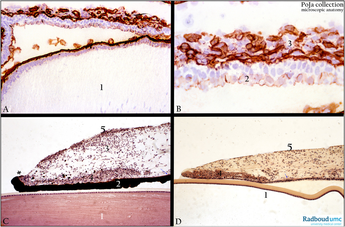
12.1.3 POJA-L2548+2550+4409+3564
Title: Iris of the eye
Description:
(A, B): Immunoperoxidase with DAB staining and anti-collagen IV antibodies, 1d postnatal, rat. (1) Lens, with a collagen IV-
positive capsule covering. The detail in (B): Collagen IV is, of course, strongly present in the loosely connective tissue (3) in
the iris, while the basement membranes of pigmented epithelial cells at the posterior site of the iris (2) are only weakly positive.
(C, D): Pupillary portion of the iris, stain Haematoxylin-Weigert, human respectively albino rabbit.
The lens (1) is covered with a single epithelial lining associated with the capsule.
The anterior iris surface (5) is a border layer consisting of fibroblasts and melanocytes.
The double epithelial layer at the posterior side is strongly pigmented (2).
The pigment ruff (* ) is close to the termination of the sphincter muscle (C, 4).
The pigmented myoepithelial cells comprise the dilator muscle that ends behind the sphincter muscle (thin arrows in C, D).
(D, 4) Is the sphincter muscle of the pupil. The posterior lining of the iris in the albino rabbit consists of a non-pigmented “outer”
epithelial layer and an “inner” pigmented myoepithelial layer. The latter layer is also called dilator muscle (thin arrow).
Keywords/Mesh: eye, iris, sphincter muscle, dilator muscle, myoepithelial cell, collagen IV, histology, POJA collection

