
13.1 POJA-L4732B+4685 Aorta (human)
13.1 POJA-L4732B+4685
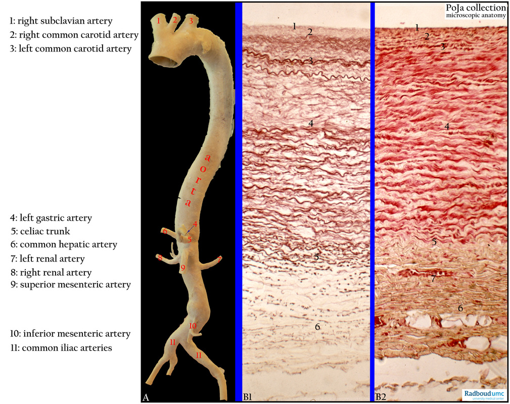
Title: Aorta (human)
Description:
(A): Aorta (isolated specimen), macroscopy. Legends in the A-frame.
(B1-B2): Aorta, orcein (B1) and azocarmine, light green (B2). (1) Endothelium, (2) subendothelial layer (1+2 = intima). (3) Internal elastic lamina or IEL (a fenestrated band of elastic fibres at the boundary of intima and media). (4) Media (mainly circular smooth muscle cells and supported by a framework of variable numbers of collagen fibres, extracellular matrix (ECM) and elastic sheaths with irregular perforations. In conducting vessels (e.g. aorta) the amount of elastic sheaths is increased and smooth muscle bundles (strain muscles) permeate between the elastic lamellae. (5) External elastic lamina or EEL might be discerned as a fenestrated layer at the boundary of media and adventitia. (6) Adventitia (loose connective tissue with short elastic fibres, collagen tissue and vasa vasorum/nervi including lymphatics), (7) vasa vasorum.
Background: In human the intima of the aorta is relatively thick compared with the thinner intima in the dog’s aorta. Thoracic aorta media contains about 50-70 lamellae and 30-40 lamellae in the abdominal aorta. The internal elastic lamina is the first lamella in the media. The amount of external tunics (adventitia) varies between human, dog and cow. Sympathetic and parasympathetic nerve plexus (nervi vasorum) are located with myelin-free processes in the adventitia. Postganglionic neurons contain free varicosities without wrapping by Schwann cells. Basal laminae of axon and smooth muscle cells are found fused.
Keywords/Mesh: cardiovascular system, vascularisation , blood vessel, aorta, elastic artery, vasa vasorum, histology, macroscopy, POJA collection