
14.3 POJA-L6309+6095 Electron micrograph of capillary and arteriole in skeletal muscle
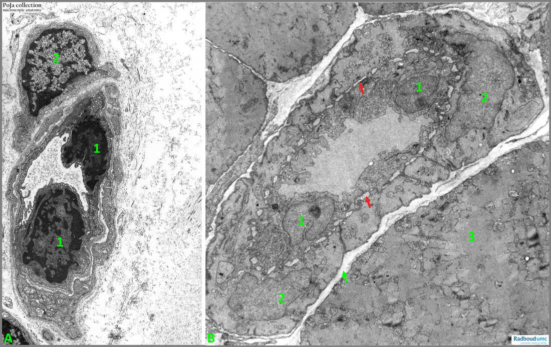
14.3 POJA-L6309+6095 Capillary and arteriole in skeletal muscle
14.3 POJA-L6309+6095 Electron micrograph of capillary and arteriole in skeletal muscle
Title: Electron micrograph of capillary and arteriole in skeletal muscle
Description:
(A): Capillary with two endothelial cells (1) and a pericyte (2) in perimysial tissue of a muscle (human). Note the numerous pinocytic vesicles in the endothelial cells.
(B): Arteriole in endomysial muscle tissue (mouse). (1): Endothelial cells. (2): surrounding smooth muscle cells. (3): Muscle fibre. Arrow points to the endomysium around the myofibre. The red arrows point to the internal elastic membrane. The same image is previously published as 13.1 POJA-L4592+4600+4632+4601+4635+4636+La0297 Arterioles.
See also:
Keywords/Mesh: locomotor system, skeletal muscle, smooth muscle, pericyte, vascularisation, capillary, arteriole, electron microscopy, POJA collection
Description:
(A): Capillary with two endothelial cells (1) and a pericyte (2) in perimysial tissue of a muscle (human). Note the numerous pinocytic vesicles in the endothelial cells.
(B): Arteriole in endomysial muscle tissue (mouse). (1): Endothelial cells. (2): surrounding smooth muscle cells. (3): Muscle fibre. Arrow points to the endomysium around the myofibre. The red arrows point to the internal elastic membrane. The same image is previously published as 13.1 POJA-L4592+4600+4632+4601+4635+4636+La0297 Arterioles.
See also:
- 13.1 POJA-L4642+La0301+4721+4626+4625 Capillaries: inner and outer lining cells
- 13.1 POJA-L2979+4643+4644+4692+4334 Ultrastructure of capillaries
Keywords/Mesh: locomotor system, skeletal muscle, smooth muscle, pericyte, vascularisation, capillary, arteriole, electron microscopy, POJA collection