
55. POJA- L2484+2483+
2485+2486.
Interstitium II of the cortex in the kidney
5.5 POJA-L2484+2483+2485+2486
Title: Interstitium II of the cortex in the human kidney
Description:
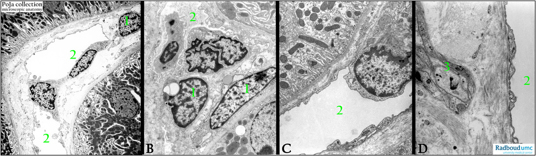
Electron microscopy. (A): Cortical interstitial cell type located between basal portions of proximal tubules.
(B): Peritubular cortical interstitial cells.
(C): Capillary apposed to proximal straight tubules (PST).
(D): Fenestrated capillary, axon and collagen. (1) Interstitial cells between renal tubules.
(2) Interstitial capillaries lined by fenestrated endothelial cells.
(3) Nerve fiber in interstitium.
Keywords/Mesh: urinary system, kidney, cortex, interstitium, myofibroblast, fenestrated capillary, histology, electron microscopy, POJA collection
Title: Interstitium II of the cortex in the human kidney
Description:
Electron microscopy. (A): Cortical interstitial cell type located between basal portions of proximal tubules.
(B): Peritubular cortical interstitial cells.
(C): Capillary apposed to proximal straight tubules (PST).
(D): Fenestrated capillary, axon and collagen. (1) Interstitial cells between renal tubules.
(2) Interstitial capillaries lined by fenestrated endothelial cells.
(3) Nerve fiber in interstitium.
Keywords/Mesh: urinary system, kidney, cortex, interstitium, myofibroblast, fenestrated capillary, histology, electron microscopy, POJA collection