9.3 POJA-L3499+3506+3498
Title: Calcitonin cells (C-cells) or parafollicular cells in thyroid gland (VII)
Description:
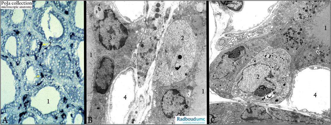
Thyroid gland. (A): Enzyme histochemistry,Gpox, glycerophosphate oxidase, pig. (B, C): Electron microscopy, gerbil.
The Gpox histochemical staining (A) reveals the presence of darkly stained parafollicular cells (2, arrows) just below the follicular
epithelial cells which line the follicle (1). The parafollicular cells (B, 2 and C, 2) contain numerous small light-grey granules (B, 2)
(probably due to granule dissolution) as well as electron-dense granules (calcitonin), but a limited amount of RER.
The follicular epithelial cells (B, 3 and C, 3) however are rich in RER and Golgi complexes due to the production of thyroglobulin.
Their electron-dense vacuoles are much larger and irregular and localized in the apex of the cell. Note also the basal-lateral
invaginations in the follicular cells (B, 3) related to the iodine pump activity. (4) Fenestrated capillaries.
(B, 5) Slender process of a macrophage.
Keywords/Mesh: thyroid gland, follicle, follicular cell, parafollicular cell, calcitonin cell, C-cell, calcitonin, thyroglobulin, histology, electron microscopy, POJA collection

