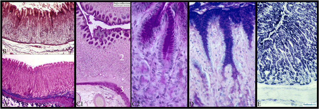
4.1.1 POJA-L3957+3958+3963+3965+3974
Title: Mucosa of fundus of the stomach (human, cat, dog, mouse)
Description: Stain: (A) Azan, human. (B) Alum hematoxylin-eosin azocarmin, human. (C1, 2) PAS, cat. (D) Alcian blue, dog. (E) Gpox, mouse.
The mucosa of the fundus consists of gastric pits with foveolae (C1-1) covered with simple columnar epithelial cells that produce mucus for protection of the cell surface against HCL and pepsinogen. The gastric glands (C1-2) are tubular and end on the lamina muscularis mucosae (C1-3). The other muscular layers are not shown here.
The azocarmin staining in (B) depicts the upper mucus producing cells as well as the parietal cells, while the lower parts of the glands with abundant chief cells are less stained.
Both the PAS staining (C1, C2) and the Alcian Blue staining (D) illustrate that the protective mucus is predominantly secreted in the area of the foveolae by the surface cells and the neck cells. The protective mucus is subsequently spread over the surface of the epithelium.
(E) Gpox staining is indicative for the mitochondrial content of the cells, excessively present in the parietal cells.
Keywords/Mesh: stomach, fundus, PAS, Alcian blue, Gpox, mucus, histology, POJA collection

