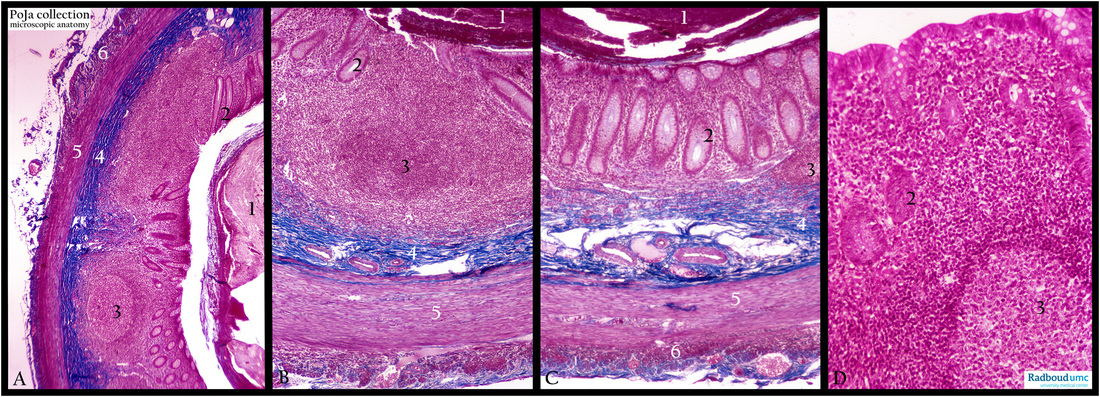
4.1.1 POJA-L-1086+4076+4077+4078
Title: Appendix (human)
Description: Stain: Azan. Survey and details of the appendix.
(1): Food debris in the lumen of the appendix.
(2): Crypts covered with simple columnar epithelium with goblet cells and enterocytes.
(3): The lamina propria and locally also the lamina muscularis mucosae is completely filled up here by lymph follicles.
(4): Tela submucosa.
(5): Inner circular muscle layer.
(6): Part of the outer longitudinal muscle layer covered with a small rim of serosa tissue.
The follicles in the appendix are part of the so called GALT, i.e. gut associated lymphoid tissue, to which also the Peyer’s patches belong. In an inflammatory process the lymphocytes can expand considerably and mask the other layers of the appendix.
Keywords/Mesh: colon, appendix, large intestine, GALT, lymphoid follicles, histology, POJA collection

