
13.1 POJA-La0301+L4600+La0296+L4588+4689+4660+4658+4678+4683 Blood and lymph vessels
13.1 POJA-La0301+L4600+La0296+L4588+4689+4660+4658+4678+4683
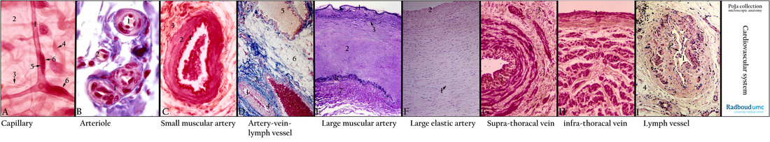
Title: Blood- and lymph vessels
Description: In this strip the individual magnifications of the light microscopy are irrelevant. The strip shows in a ‘bird’s-eye ‘view the sequence of representative types of vessels.
(A): Capillaries (mesenterium), hematoxylin-Weigert, frog. Thin capillary (1) in the areolar loose connective tissue (2). Thin elastic fibres (arrow 3) and coarse collagen fibres (arrow 4). Capillary (endothelial cell, arrow 5) is surrounded by pericytes (arrow 6), these contractile cells contain i.a. actin and regulate the diameter of a capillary. Upon stimulation pericytes are able to phagocytise.
(B): Arterioles and capillaries (omentum), Azan, human. (1) Arterioles, note one or few SMC in the tunica media. (2) Capillary.
(C): Muscular artery (small artery in omentum), orcein-azocarmine, human. (1) Well-developed internal elastic membrane. (2) Fine branching and distribution of thin elastic fibres in media. (3) Adventitia with discontinuous external elastic membrane and few elastic fibres.
(D): Arteries/vein/lymph vessel (subcutis of skin), Azan, human. Artery (1) (with erythrocytes) showing a thicker media with circular as well as partly cross-sectioned smooth muscle cells (4). (2) Adventitia artery/vein. (3) Vein. (5) Dilated lymph vessel with a thin wall. (6) Adipose tissue of the subcutis.
(E): Popliteal artery (muscle biopsy), toluidine blue-stained 1 micrometer plastic section, human. In this biopsy the intima is thin and closely followed by a distinct internal elastic lamina (1). In the upper 1/3 of the media (2) increase of elastic fibres (arrow 3) and deeper the circular and longitudinal layers of smooth muscle cells (SMC). The boundary with the adventitia (7) is only marked by circular concentrations of elastic fibres (8).
(F): Common carotid artery, HE, human. When contrast is enhanced by closing the diaphragm aperture numerous unstained elastic membranes in the media appear distinct as tortuous unstained lamellae (arrow 1). Intima (2).
(G): Mid-sized vein of upper body half (branch of jugular vein), resorcin-fuchsin and light green, human. This vein contains thin longitudinal arranged smooth muscle cells (SMC) in the intima (1) and irregularly circular arranged SMC bundles in the media (2) and the adventitia (3). Bundles of SMC in the adventitia are absent because hydrostatic pressure contributes to the down flow of blood circulation. This vein possesses a thinner wall compared to veins of the lower body half that need their adventitial muscles as the propelling-force for the upwards flow.
(H): Mid-sized vein of lower body half (branch of femoral vein), resorcin-fuchsin and light green, human. Small intima (1). Hardly developed media with some circular SMC. Well-developed adventitia (2) with longitudinal arranged bundles of SMC. Along the whole vein the wall appears irregular thick. The adventitial muscles are the propelling-force of the blood flow in direction of the heart.
(I): Lymphatic vessel (mesentery), HE, human. At low magnification lumen of lymphatic vessels (1) with valves (arrow 2), note irregular bundles smooth muscle cells in the wall (3) and adipose tissue (4).
Keywords/Mesh: cardiovascular system, vascularisation, lymphatic system, capillary, arteriole, artery, muscular artery, elastic artery, vein, lymphatic vessel, histology, POJA collection