6.1 POJA-L2942+La0187+2687+2689+2690+La0194
Title: Spermatogenesis in testes
Description:
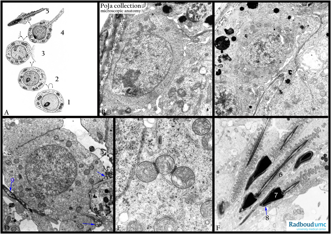
(A) Electron microscopy scheme. (B-F) Electron microscopy, gerbil.
(A): The spermatogenesis (scheme A) comprises initially a spermatogonium of type A (stem cell, A, 1) all arranged as a basal layer in the seminiferous tubules. After cell division some cells further differentiate into primary spermatocytes (2). After some more meiotic divisions these cells duplicate into secondary spermatocytes (3). These cells after some mitosis further differentiate into spermatids (4) and finally into spermatozoa (5).
(B): Spermatogonium.
(C): Primary spermatocyte.
(D, E): Spermatid. In (D, 9) slender darkly stained extensions of a Sertoli cell are visible.
(F): Maturing spermatozoa.
(E, F): From (E-F) the mitochondria (F, 6) will be arranged spirally around the small longitudinal axis, just below the head (F, 7). The head in fact is no more than a strongly condensed nucleus covered by the acrosome (F, 8). Note that the maturing spermatozoa are still sticking into the cytoplasm of the Sertoli cell. In (D, 9) slender darkly stained extensions of a Sertoli cell are visible.
Keywords/Mesh: testis, seminiferous tubule, spermatogenesis, spermiogenesis, spermatogonium, histology, electron microscopy, POJA collection

