
5.4.3 POJA-L2371+2312+
2436+2397+4256+4259.
Proximal convoluted tubules (PCT) and proximal straight tubules (PST) (II) in the human kidney
5.4.3 POJA-L2371+2312+2436+2397+4256+4259
Title: Proximal convoluted tubules (PCT) and proximal straight tubules (PST) (II) in the human kidney
Description:
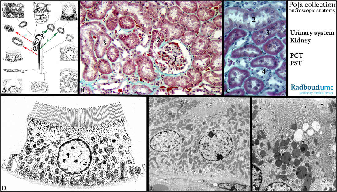
(A): Scheme nephron. (Zoom in). The red arrows (1 and 2) point to the structure of the proximal convoluted tubules (1, PCT) with
ample microvilli and the proximal straight tubule (2, PST), while the green arrows (7 and 5) point to the distal straight tubules (5, DST),
with hardly developed microvilli, and the distal convoluted tubule (7, DCT).
(B): Stain trichrome Goldner, glomerulus (1) showing the direct transition at the urinary pole into the proximal convoluted tubule (2).
The distal tubules are stained slightly lighter due to the lower amount of mitochondria and microvilli.
(C): Stain trichrome Masson showing (3) PCT and PST (2), (4) distal tubule (DST).
(D): Electron microscopy scheme of a proximal tubule cell displaying numerous apical microvilli and endocytosis vesicles.
The basal membrane has deep invaginations between which the mitochondria are arranged.
(E, F): Electron micrographs of the microvillar structure on the apical side of the cells of a proximal tubule (PCT).
Note the mitochondrial arrangement and the numerous lysosomal structures. The mitochondria provide the energy for the active transport (ATPase) of Na+ ions followed by passive diffusion of Cl- ions and water. Proteins (endocytosis) and amino acids are also reabsorbed from the glomerular filtrate.
Keywords/Mesh: urinary system, kidney, glomerulus, proximal tubule, distal tubule, histology, electron microscopy, POA collection
Title: Proximal convoluted tubules (PCT) and proximal straight tubules (PST) (II) in the human kidney
Description:
(A): Scheme nephron. (Zoom in). The red arrows (1 and 2) point to the structure of the proximal convoluted tubules (1, PCT) with
ample microvilli and the proximal straight tubule (2, PST), while the green arrows (7 and 5) point to the distal straight tubules (5, DST),
with hardly developed microvilli, and the distal convoluted tubule (7, DCT).
(B): Stain trichrome Goldner, glomerulus (1) showing the direct transition at the urinary pole into the proximal convoluted tubule (2).
The distal tubules are stained slightly lighter due to the lower amount of mitochondria and microvilli.
(C): Stain trichrome Masson showing (3) PCT and PST (2), (4) distal tubule (DST).
(D): Electron microscopy scheme of a proximal tubule cell displaying numerous apical microvilli and endocytosis vesicles.
The basal membrane has deep invaginations between which the mitochondria are arranged.
(E, F): Electron micrographs of the microvillar structure on the apical side of the cells of a proximal tubule (PCT).
Note the mitochondrial arrangement and the numerous lysosomal structures. The mitochondria provide the energy for the active transport (ATPase) of Na+ ions followed by passive diffusion of Cl- ions and water. Proteins (endocytosis) and amino acids are also reabsorbed from the glomerular filtrate.
Keywords/Mesh: urinary system, kidney, glomerulus, proximal tubule, distal tubule, histology, electron microscopy, POA collection