
12.2.2 POJA-L3396+3391+
3395+3392+3393
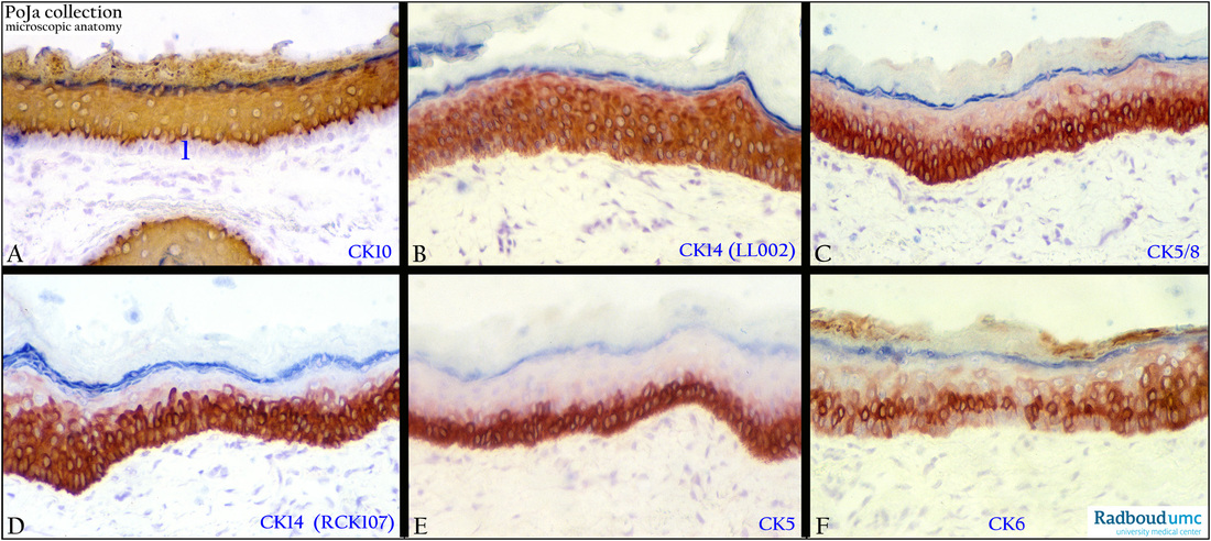
Keratin expression pattern in epidermis of the external auditory meatus in human ear
12.2.2 POJA-L3396+3391+3395+3392+3393
Title: Keratin expression pattern in epidermis of the external auditory meatus in human ear
Description:
Immunoperoxidase staining with DAB and monoclonal antibodies against different types of cytokeratin, counterstained with Haematoxylin. Note that the stratum granulosum is identifiable by the blue-stained keratohyalin granules.
(A): Anti-keratin 10 antibody (RKSE60). With exception of the basal layer (1) all epidermal cell layers are stained positively.
(B): Anti-keratin 14 antibodies (LL002). Expression remains limited to the stratum Malpighi (= stratum basale or germinativum + stratum spinosum).
(C): Anti-keratin 5/8 antibodies (RCK102). Stratum basale and 2-3 layers of the stratum spinosum above are stained positively, the rest upwards loses its expression.
(D): Anti-keratin 14 antibodies (RCK107), showing similar expression as cytokeratin 5/8.
(E): Anti-keratin 5 antibodies (AE14). A more strictly expression limited to the stratum basale cell layer and the one layer above it.
Upper layers have switched off the expression.
(F): Anti-keratin 6 antibodies (KA12). Limited and miscellaneous expression throughout the epidermis, but mainly in the suprabasal layers (youngest stratum spinosum cells). There is variable cytokeratin 6 activity in the basale or stratum germinativum.
Background:
- With exception of the basal layer cytokeratin (Ck) 10 is a cytokeratin marker for the rest of the epidermis including the corneocytes.
- Ck 5 (E) and Ck 14 (D) are the major proteins of mitotically active basal keratinocytes. They are replaced by Ck 1 (not shown here) and
Ck 10 when these cells migrate toward the stratum spinosum. The latter Cks 1 and 10 provide mechanical integrity to the cells and they also participate in signaling processes.
- Note that Ck 5 and Ck 14 are absent in the non-dividing stratum granulosum (where keratohyalin formation, production of fillagrin occurs) and the stratum corneum ( where production of cornified cell envelopes and outer multilamellar lipid envelopes takes place).
- During wound healing (hyperproliferative stimuli) or in some carcinomas the expression of Cks 5 and 14 remain active while Cks 1 and
10 are temporarily reduced.
- The Ck 6 becomes upregulated again in hyperproliferative epidermis in certain conditions, hence the variable reactivity in (F).
- Some dominant mutations weaken the cytokeratin proteins 5 or 14, resulting in a tendency to rupture of cell layers which than separate - from each other causing accumulation of fluid between them leading to blister formation in the skin (epidermolysis bullosa simplex).
Keywords/Mesh: external ear, epidermis, cytokeratin, histology, POJA collection