12.1.2 POJA-L4391+2532+2533+4392
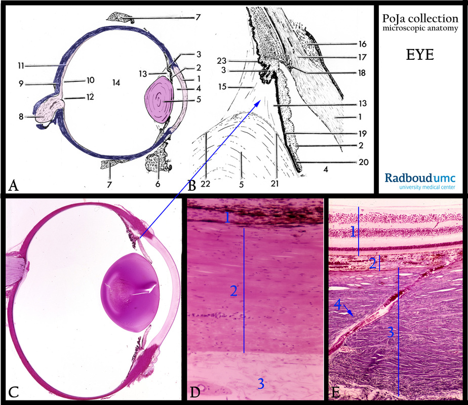
Title: Survey of the structure of the eyeball (bulbus oculi)
Description:
(A, B): Scheme of the eye and as detail the ciliary body in (B), human.
(1) Cornea.
(2) Iris.
(3) Ciliary body.
(4) Anterior chamber of the eye.
(5) Lens cells.
(6) Palpebral conjunctiva.
(7) Extraocular muscle.
(8) Optic nerve.
(9) Sclera.
(10) Fovea centralis (macula).
(11) Retina (optical part).
(12) Optic disc.
(13) Posterior chamber.
(14) Vitreous body.
(15) Zonular fibres.
(16) Bulbar conjunctiva.
(17) Limbus of cornea.
(18) Trabecular meshwork (aqueous humor).
(19) Ciliary portion of iris.
(20) Sphincter muscle at the pupillary portion of iris.
(21) Equatorial capsule with lens epithelium.
(22) Lens capsule.
(23) Ciliary processes.
(C): Survey of the bulbus oculi, stain Haematoxylin-eosin, human.
(D): Three layers of the sclera, stain Haematoxylin-eosin, human. (1) Inner layer of sclera or lamina fusca with pigmented cells.
(2) Middle layer (substantia propria) of sclera. (3) Outer layer of sclera or episclera.
(E): Emissarium, stain Azan, human. (1) Inner tunic (retina). (2) Middle tunic (vascular layer). (3) Outer tunic (sclera without cornea).
(4, arrow) So-called emissarium (groups of anterior and posterior channels with blood vessels/nerves through the sclera towards the middle tunic).
Keywords/Mesh: eye, cornea, iris, ciliary body, retina, sclera, emissarium, histology, POJA collection

