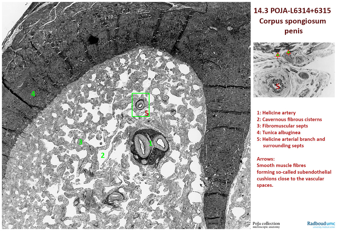
14.3 POJA-L6314+6315 Electron micrograph of smooth muscles in corpus spongiosum of the penis I (human)
a 14.3 POJA-L6314+6315 Electron micrograph of smooth muscles in corpus spongiosum of the penis I (human)
Title: Electron micrograph of smooth muscles in corpus spongiosum of the penis I (human)
Description:
The penis consists of the paired corpora cavernosa and the corpus spongiosum of the urethra-seminal duct. They are covered by the tunica albuginea (4), a tough and dense connective tissue with fibroelastic properties. The cavernous spaces are formed by fibro-collagenous strengths or stroma supported by smooth muscle cells to regulate the storage and blood flow in the cisternal spaces or vascular canals.
See also:
During erection the helicine arteries dilate and the vascular cisterns increase in size by filling with blood from these aforementioned arteries.
Keywords/Mesh: locomotor system, penis, erection, corpus spongiosum, artery, smooth muscle, helicine artery, histology, POJA collection
Description:
The penis consists of the paired corpora cavernosa and the corpus spongiosum of the urethra-seminal duct. They are covered by the tunica albuginea (4), a tough and dense connective tissue with fibroelastic properties. The cavernous spaces are formed by fibro-collagenous strengths or stroma supported by smooth muscle cells to regulate the storage and blood flow in the cisternal spaces or vascular canals.
See also:
- 14.3 POJA-L6307+6308 Branching myocytes in the corpus spongiosum of the penis II
During erection the helicine arteries dilate and the vascular cisterns increase in size by filling with blood from these aforementioned arteries.
Keywords/Mesh: locomotor system, penis, erection, corpus spongiosum, artery, smooth muscle, helicine artery, histology, POJA collection

