
2.3 POJA-L1014. Lymph node (rat)
2.3 POJA-L1014
Title: Lymph node (rat)
Description: Electron microscopy.
In the paracortical area (inner cortex) of a lymph node interdigitating cells (1) are found (so-called APC, IDC). They show branched projections (3) in between the closely apposed surrounding T lymphocytes (2). In their cytoplasm lysosomal inclusions of different sizes and content (*) might be present and sometimes tubulovesicular profiles can be discerned. (4) Macrophages with many small lysosomal structures.
Keywords/Mesh: lymphatic tissue, lymph node, reticular tissue, interdigitating cell, antigen-presenting cell, lysosome, histology, electron microscopy, POJA collection
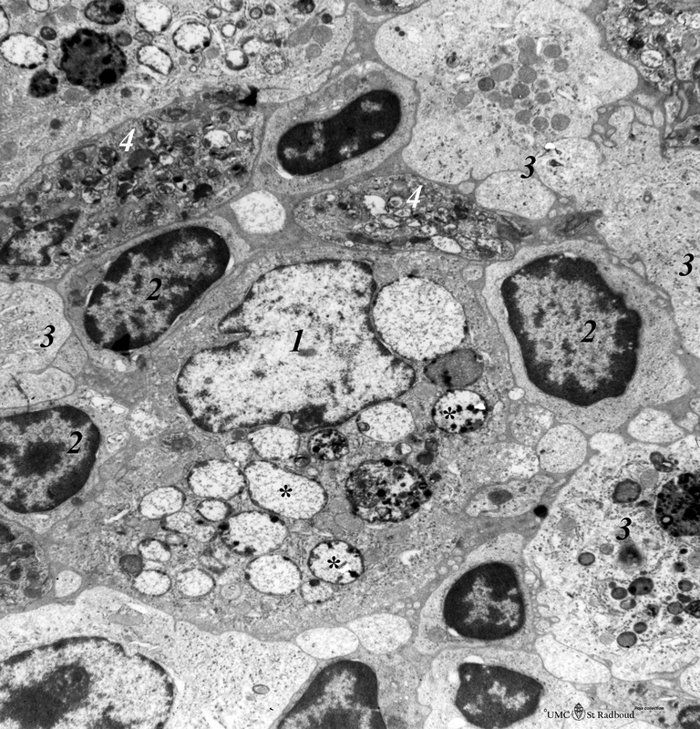