
11.6 POJA-L3866+3369+
3867+3868.
Pathology of multiple sclerosis
11.6 POJA-L3866+3369+3867+3868
Title: Pathology of multiple sclerosis
Description:
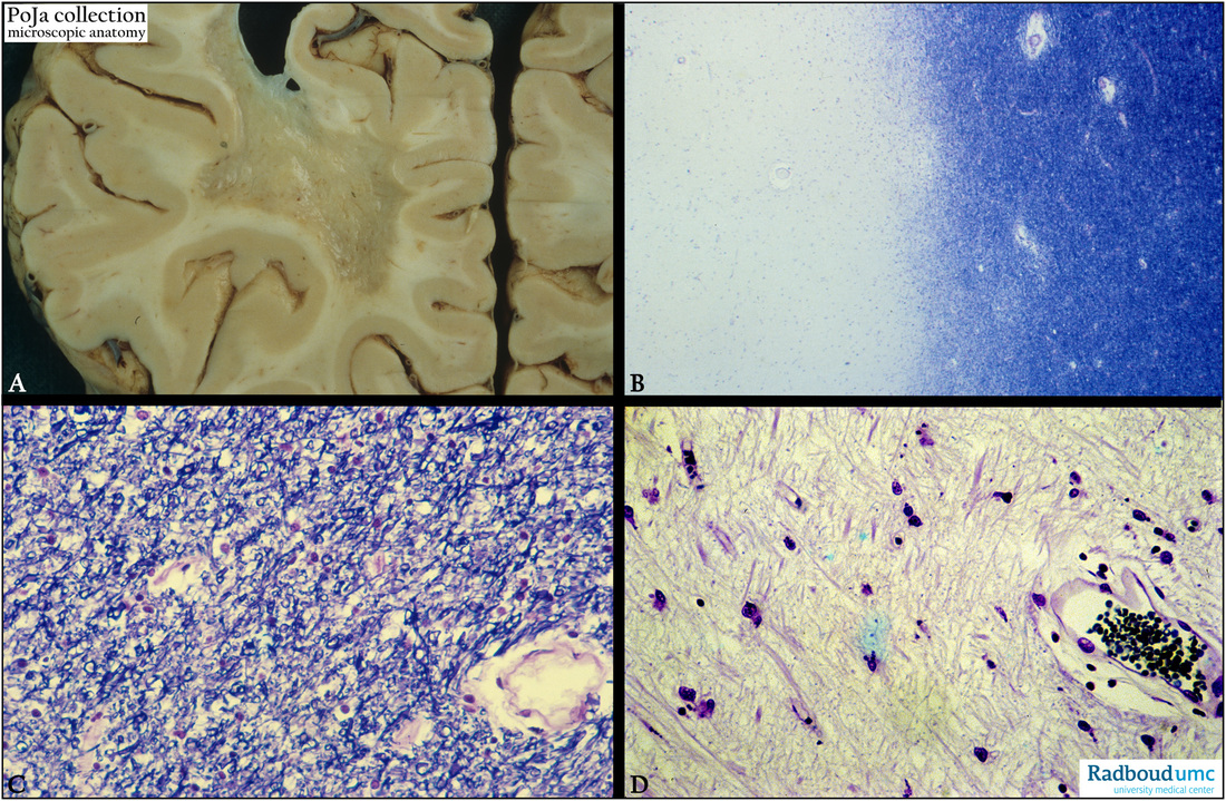
(A): Macroscopy of multiple sclerosis (MS) as example of a demyelinating disease in the cerebrum, revealing a slimy gelatinous gray-white appearance of the former white substance, human.
(By courtesy of P. Wesseling MD PhD, Head Neuropathology, Department of Pathology, Radboud university medical center, Nijmegen, The Netherlands).
(B): Kluver-Barrera staining (with Luxol fast blue for lipoproteins of myelin) of an affected area (plaque) of MS illustrates the dissolution
of the myelin (blue) in the decolorized white matter, while the blue-stained part with myelin is still not affected (human).
Note the presence of the small blood vessels.
(C): Kluver-Barrera staining of an unaffected part of the cerebral medulla with normal glia cells, blood vessels, human.
(D): Kluver-Barrera staining of a demyelinated cerebrum area showing a considerable dissolution of the glia tissue, human.
MS primarily affects the demyelination of the white matter in the periventricular distribution. However gray matter demyelination may occur as well in the motor cortex, cerebellum, spinal cord, junction of cortex and white matter, and at the edges of the lateral ventricles.
Bottom right shows an arteriole with paravascular spaces (Vichow-Robin) belonging to the glymphatic pathway for exchange of solutes as well as a para-venous clearance route for removal of brain interstitial fluid. (See also: Glymphatics POJA-L4750)
Keywords/Mesh: nervous tissue, cerebrum, multiple sclerosis, demyelination, pathology, histology, POJA collection
Title: Pathology of multiple sclerosis
Description:
(A): Macroscopy of multiple sclerosis (MS) as example of a demyelinating disease in the cerebrum, revealing a slimy gelatinous gray-white appearance of the former white substance, human.
(By courtesy of P. Wesseling MD PhD, Head Neuropathology, Department of Pathology, Radboud university medical center, Nijmegen, The Netherlands).
(B): Kluver-Barrera staining (with Luxol fast blue for lipoproteins of myelin) of an affected area (plaque) of MS illustrates the dissolution
of the myelin (blue) in the decolorized white matter, while the blue-stained part with myelin is still not affected (human).
Note the presence of the small blood vessels.
(C): Kluver-Barrera staining of an unaffected part of the cerebral medulla with normal glia cells, blood vessels, human.
(D): Kluver-Barrera staining of a demyelinated cerebrum area showing a considerable dissolution of the glia tissue, human.
MS primarily affects the demyelination of the white matter in the periventricular distribution. However gray matter demyelination may occur as well in the motor cortex, cerebellum, spinal cord, junction of cortex and white matter, and at the edges of the lateral ventricles.
Bottom right shows an arteriole with paravascular spaces (Vichow-Robin) belonging to the glymphatic pathway for exchange of solutes as well as a para-venous clearance route for removal of brain interstitial fluid. (See also: Glymphatics POJA-L4750)
Keywords/Mesh: nervous tissue, cerebrum, multiple sclerosis, demyelination, pathology, histology, POJA collection