
7.1 POJA-L1628+1740+1742+1333. Ultrastructure of follicles in ovary (gerbil, rabbit)
7.1 POJA-L1628+1740+1742+1333
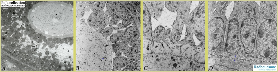
Title: Ultrastructure of follicles in ovary (gerbil, rabbit)
Description: Electron microscopy (gerbil, rabbit).
(A): Gerbil: low magnification shows part of multilaminar secondary follicle (late type 4) with antrum. Vesicular nucleus (without nucleolus) (1) and perinuclear accumulation of mitochondria (part of Balbiani body) (2). Peripherally electron-lucent vacuoles in ooplasm close to the zona pellucida (3). Corona radiata cells (4) and small antrum filled with granular material, free-floating intact granulosa cells as well as apoptotic granulosa cells (5).
(B): Rabbit: secondary follicle with ooplasm and dispersed mitochondria, small electron-dense cortical granules (→). A well developed zona pellucida (*) is already present between oocyte and two-layered mural granulosa cells. Hardly detectable at light microscopical level ultrastructurally distinct intercellular spaces with liquor are already present between granulosa cell cytoplasms.
(C): Rabbit: At higher magnification of a similar follicle the peripheral rim of ooplasm contains numerous small vesicles, electron-dense cortical granules as well as dense lamellated structures. Within the broad zone of basal lamina-like zona pellucida (*) oocytic microvilli are in close contact with interzonal villi from feet-like extensions of granulosa cells. Their apices are junctioned together and intercellular spaces with liquor are present.
(D): Rabbit: the same follicle as in (C) shows mural granulosa cells bordered by a basal lamina reinforced with collagen fibers (basement membrane or membrana vitrea at light microscopical level) (è-ç). Their nuclei are palisade-like arranged and lipoid droplets (6) are already present. At bottom side layering of fibroblast-like flattened inner theca cells (7) containing long profiles of rough endoplasmic reticulum.
Background: Oocytes of different species contain Balbiani bodies with a heterogeneous morphology and composition. Generally this transient structure is located juxtanuclearly and consists of a.o. accumulation of mitochondria, Golgi areas, endoplasmic reticulum, lipids, fibrogranular material, RNAs. It is thought to function in the organization and transport of organelles and RNAs to specific regions of the ooplasm. The zona pellucida plays an important role in the fertilization process; it is composed of a.o. glycoproteins ZPGp1, ZPGp2, ZPGp3 and ZPGp4. It is known that capacitation (occurring as sperm remains one up to six hours in the female genital tract) includes the process of loss of spermatozoal glycocalyx as well as vesiculation of the acrosome of spermatozoon. In the binding process of spermatozoa oligosaccharides linked to ZPGp3 interact with sperm receptors. It appears that only acrosome-reacted spermatozoon can interact with ZPGp3. The cortical reaction refers to the exocytosis of cortical granules (produced by Golgi areas) discharging their contents (mixtures of proteases) in the zona pellucida shortly after sperm-oocyte fusion. Proteases alter the pellucidal structure resulting in a specific transformation of the zona pellucida that penetration by other capacitated spermatozoa (polyspermy) is prevented (so-called zona reaction).
Keywords/Mesh: female genitalia, ovary, ovarian follicle, oocyte, zona pellucida, granulosa cells, electron mcroscopy, histology, POJA collection