
2.2 POJA-L976B+974. Scheme electron microscopy of the border of secondary nodule/red pulp in spleen
2.2 POJA-L976B+974
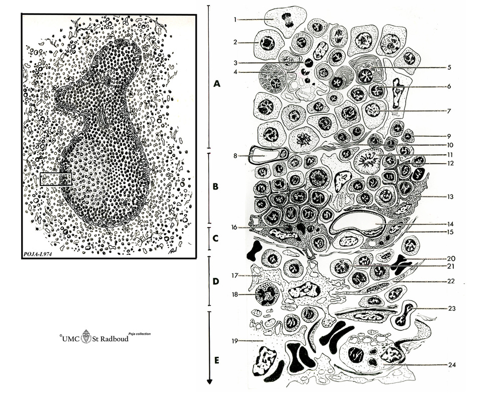
Title: Scheme electron microscopy of the border of secondary nodule/red pulp in spleen
Description:
The left figure POJA-L974 shows a scheme of histological impression of a survey of a secondary splenic follicle or nodule. The rectangle is enlarged in the right figure POJA-L976B and shows the following elements:
(A): Germinal centre
(B): Mantle zone
(C): Dendritic cell area
(D): Marginal zone
(E): Red pulp
Background: The marginal zone contains macrophages that are particularly efficient at trapping polysaccharide antigens. Such antigens may persist for prolonged periods on the surfaces of marginal zone macrophages, where they are recognized by specific B cells, or they may be transported into the follicle. The marginal zone macrophages and the metallophilic macrophages, however, although essential for trapping antigen, are dispensable for Ag presentation. The metallophilic marginal zone macrophages are more involved in the production of Interferon-α/β than in antigen-presenting. They also miss the expression of MHC-class-II and CD11c. The marginal zone macrophages are also involved in the transient retention of B cells on their move to the follicle centre. It is suggested that the marginal zone macrophages in the spleen are analogs of HEV’s (high endothelial venules) in the lymph node, forming the port of entry of lymphocytes into the white pulp of the spleen.
Keywords/Mesh: lymphatic tissue, spleen, white pulp, red pulp, lymphatic nodule, germinal centre, reticular tissue, marginal zone, mantle zone, dendritic APC, histology, electron microscopy, POJA collection.
Title: Scheme electron microscopy of the border of secondary nodule/red pulp in spleen
Description:
The left figure POJA-L974 shows a scheme of histological impression of a survey of a secondary splenic follicle or nodule. The rectangle is enlarged in the right figure POJA-L976B and shows the following elements:
(A): Germinal centre
(B): Mantle zone
(C): Dendritic cell area
(D): Marginal zone
(E): Red pulp
- mitosis of lymphoblast
- lymphoblast
- macrophage with ‘tingible bodies’
- proplasmacyte
- mature plasma cell
- maturing B lymphocyte
- large lymphocyte
- capillary
- small lymphocyte
- reticular fibres
- mitotic lymphoblast in mantle zone
- projection of branching (metallophilic) dendritic antigen-presenting cell (APC)
- (metallophilic) dendritic APC
- capillary of anastomosing vascular channels
- reticular fibres
- macrophage with phagocytised material
- macrophage in marginal zone
- neutrophilic granulocyte
- macrophage in red pulp
- erythrocyte in marginal sinus
- monocyte
- (flibroblastic) reticular cell
- diapedesis of lymphocyte through wall of perimarginal sinus
- phagocytising sinus endothelial cell
Background: The marginal zone contains macrophages that are particularly efficient at trapping polysaccharide antigens. Such antigens may persist for prolonged periods on the surfaces of marginal zone macrophages, where they are recognized by specific B cells, or they may be transported into the follicle. The marginal zone macrophages and the metallophilic macrophages, however, although essential for trapping antigen, are dispensable for Ag presentation. The metallophilic marginal zone macrophages are more involved in the production of Interferon-α/β than in antigen-presenting. They also miss the expression of MHC-class-II and CD11c. The marginal zone macrophages are also involved in the transient retention of B cells on their move to the follicle centre. It is suggested that the marginal zone macrophages in the spleen are analogs of HEV’s (high endothelial venules) in the lymph node, forming the port of entry of lymphocytes into the white pulp of the spleen.
Keywords/Mesh: lymphatic tissue, spleen, white pulp, red pulp, lymphatic nodule, germinal centre, reticular tissue, marginal zone, mantle zone, dendritic APC, histology, electron microscopy, POJA collection.