16.1.3 POJA-L7083+7084 Blood vessels in compact bone (cortical bone) 4
16.1.3 POJA-L7083+7084 Blood vessels in compact bone (cortical bone) 4
Title: Blood vessels in compact bone (cortical bone) 4
Description:
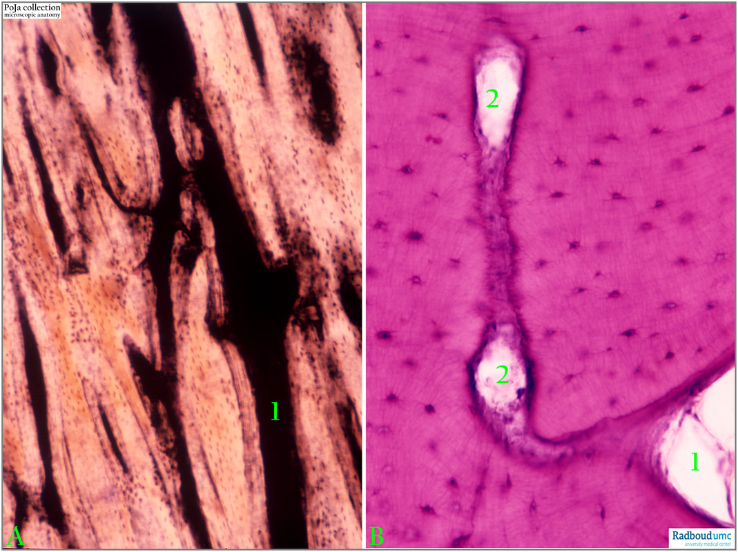
(A): The vascular system in compact bone has been visualized by injecting Indian ink staining black the Haversian canals (1) where blood vessels and capillaries are located.
(B): Haematoxylin-eosin stain of compact bone with Haversian canal (1) and osteocytes.
The Haversian canals do communicate with the perforating Volkmann’s canaliculi (2) that are running transversely to the Haversian system.
Background:
Different types of bone show a variety of bood supply, but it is known that bones containing red bone marrow are very well vascularised. In long bones the diaphyseal nutrient artery passes oblique through the cortical bone. The metaphyseal and epiphyseal arteries are small ones supplying the ends of the bones, they originate from adjacent joint arteries and terminate in the bone marrow. The extensive network of periosteal arteries are responsible for the outer layers of the cortical bone and connected to the general body circulation.
A new vascular network is discovered i.e. the so-called transcortical vessels (TCV) that can be either arterioles or venules traversing perpendicularly across the entire length of the cortical bone to form a direct connection between the endosteal and periosteal ciculations. These TCVs effectively transport more than 80% of arterial and 60% of venous blood flow in long bones. It is assumed that TCVs form the mainstay of blood circulation and contribute to a fully functional closed circulatory system. This system of vessels is used by bone marrow immune cells to reach the bloodstream and in case of an inflammatory disease e.g. arthritis the immune cells very quickly reach the source of inflammation (A network of trans-cortical capillaries as mainstay for blood circulation in long bones. (Grüneboom, A. et al. Nature Metabolism 2019 DOI:10.1038/s42255-018-0016-5).
See also:
Keywords/Mesh: locomotor system, bone, compact bone, osteon, Haversian canal, Volkmann’s canal, Indian ink, osteocyte, histology, POJA collection
Title: Blood vessels in compact bone (cortical bone) 4
Description:
(A): The vascular system in compact bone has been visualized by injecting Indian ink staining black the Haversian canals (1) where blood vessels and capillaries are located.
(B): Haematoxylin-eosin stain of compact bone with Haversian canal (1) and osteocytes.
The Haversian canals do communicate with the perforating Volkmann’s canaliculi (2) that are running transversely to the Haversian system.
Background:
Different types of bone show a variety of bood supply, but it is known that bones containing red bone marrow are very well vascularised. In long bones the diaphyseal nutrient artery passes oblique through the cortical bone. The metaphyseal and epiphyseal arteries are small ones supplying the ends of the bones, they originate from adjacent joint arteries and terminate in the bone marrow. The extensive network of periosteal arteries are responsible for the outer layers of the cortical bone and connected to the general body circulation.
A new vascular network is discovered i.e. the so-called transcortical vessels (TCV) that can be either arterioles or venules traversing perpendicularly across the entire length of the cortical bone to form a direct connection between the endosteal and periosteal ciculations. These TCVs effectively transport more than 80% of arterial and 60% of venous blood flow in long bones. It is assumed that TCVs form the mainstay of blood circulation and contribute to a fully functional closed circulatory system. This system of vessels is used by bone marrow immune cells to reach the bloodstream and in case of an inflammatory disease e.g. arthritis the immune cells very quickly reach the source of inflammation (A network of trans-cortical capillaries as mainstay for blood circulation in long bones. (Grüneboom, A. et al. Nature Metabolism 2019 DOI:10.1038/s42255-018-0016-5).
See also:
- 16.1.3 POJA-L7091+7073+7075+7094 Compact bone (cortical bone) with osteons 1
- 16.0 POJA-L7079+7080+7082+7081 Compact bone (cortical bone) with osteons 2
- 16.1.3 POJA-L7085+7077+7078 Compact bone (cortical bone) with osteons and interstitial lamellae 3
- 16.1.3 POJA-L7114+7112+7093+7092 Remodeling of compact bone (cortical bone) 5
- 16.1.3 POJA-L7087+7088 Partially burned human bone specimen
- 16.1.3 POJA-L7090+7076 Bone: osteons in polarisation microscope
Keywords/Mesh: locomotor system, bone, compact bone, osteon, Haversian canal, Volkmann’s canal, Indian ink, osteocyte, histology, POJA collection

