13.1 POJA-L4736+4525+4544+4546
Title: Trabecula’s and valve of the heart
Description:
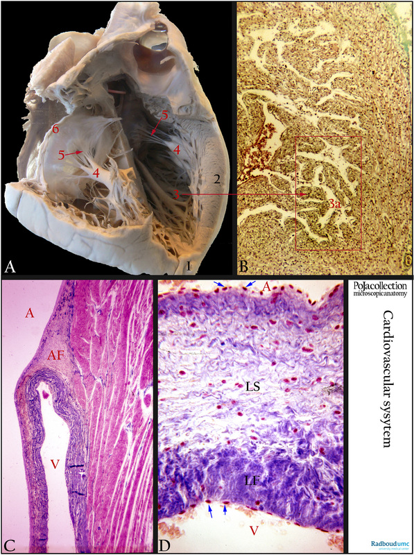
(A): Macroscopy of left side of heart, human. Apex cordis (1), thick left ventricle wall (2), trabeculae carneae (3), papillary muscle (4), chordae tendineae (arrow 5), atrioventricular valve leaflet (6).
(B): Heart, Trichrome, fetus human. Transversal section of ventricle with lumen (erythrocytes) near the apex. The cavity (3a) of the ventricle contains a lining network of irregular bundles of muscle (3) which are attached to the solid portion of the wall at their ends and run over the inner surface of this cavity. These muscular ridges are covered with endocardium and are called trabeculae carneae or Rathke’s bundles.
(C): Heart, ventricle, Azan, human. The atrioventricular valve (mitral valve) is attached to the fibrous skeleton of the heart. It consists of connective tissue located between the atria and ventricles i.e. the annulus fibrosus (AF). The valve is an endocardial fold composed of a kind of fibroelastic connective tissue without any vascularisation. In the cusp smooth muscle cells are found dispersedly located at the site of the atrium (A). At the site of the ventricle (V) the amount of dense connective tissue (blue) increases.
(D): Heart, mitral valve, Azan, human. Being an endocardial fold of fibroelastic-like connective tissue the valve itself is not vascularized. The side of the mitral leaflet facing the atrium consists of a lamina spongiosa (LS) with subendocardially loosely arranged connective tissue and nerve fibres. Few smooth muscle cells might be present at this site. The side facing the ventricle has a lamina fibrosa with dense arranged connective tissue (blue, LF). (blue arrows) Endothelial lining of the endocardium.
Background: Valves are located at the openings between the chambers and between the chambers and arteries leaving the heart. They are smooth endocardial tissue folds controlling blood flow direction within and out of the heart. Their outer margins are attached to the fibrous skeleton (annulus fibrosus) that is located between atria and ventricles. The skeleton encircles and connects each of the four valve openings and consists of dense connective tissue. Atrioventricular valves (AVvs) require contraction of papillary muscles for closing.
Further reading is referred to (Halaney, D.L. et al. in Heart Failure and Cardiomyopathies 2015); (www.idiotsguides.com/education/biology/anatomy-101-heart); Misfeld M, Sievers H-H. Heart valve macro- and microstructure. Philosophical Transactions of the Royal Society B: Biological Sciences. 2007;362(1484):1421-1436. doi:10.1098/rstb.2007.2125. http://www.ncbi.nlm.nih.gov/pmc/articles/PMC2440405/.
Keywords/Mesh: cardiovascular system, heart, atrium, ventricle, mitral valve, papillary muscle, chorda tendinae, annulus fibrosus, myocardiocyte, cardiocyte, histology, POJA collection

