
10.2 POJA-L2131+2148+2150+
2151+2152+2153+2989.
Melanocyte in the epidermis
10.2 POJA-L2131+2148+2150+2151+2152+2153+2989
Title: Melanocyte in the epidermis
Description:
(A): Skin snout, stain PAS, bovine. The pigmented dark brown keratinocytes are located in the lower half of the stratum spinosum.
The pigment is packed above the nucleus at the ‘sunny’ side of the cell.
(B): Skin arm, immunoperoxidase staining with AEC and NKI-beteb antibodies, human. These antibodies are directed against
melanin-associated antigen and used as an early differentiation marker. The melanocytes (reddish-stained) are located in
the lower layers of the epidermis and in the dermis.
(C, D, E - G): Electron microscopy scheme (C) and skin arm, electron micrograph of the electron-light melanocyte (D), human.
The enzyme tyrosinase is produced in the RER and collected in Golgi vesicles (in E), called primary melanosomes (stage 1) (1).
Melanosome (stage 2) (2) contains filamentous helical tyrosinase molecules. The electron-dense melanin is now produced and
accumulates in the released vesicles (stages 3 in G) and (stage 4 in F) and transported to the elongated slender extensions
of the cell (F). The next step includes the cytocrine secretion and passing over into the neighboring keratinocytes.
Cytoplasmic extensions of one melanocyte establish contact with ca 36 keratinocytes creating an epidermal-melanin unit.
Upon UV radiation the keratinocytes secrete many paracrine factors that influence on melanocytes, such as bFGF, ET-1,
IL-1a/1b, ACTH, and more, affecting the melanogenesis and proliferation.
Keywords/Mesh: skin, epidermis, keratinocyte, melanocyte, tyrosinase, melanosome, melanin, histology, electron microscope,
POJA collection
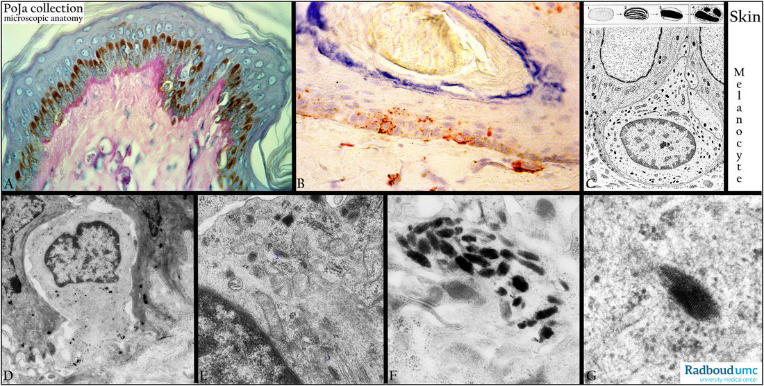