
4.2.1 POJA-L3669+3676. Liver sinusoids injected with peroxidase, (rat)
4.2.1 POJA-L3669+3676
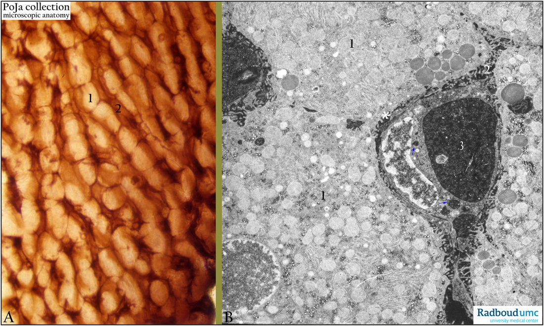
Title: Liver sinusoids injected with peroxidase, (rat)
Description: Stain: (A) One minute after injection of peroxidase and stained with DAB. (B) Electron micrograph after injection of peroxidase (DAB-stained), not contrasted.
The liver parenchymal cells (A, 1) are lucent and unstained while the sinusoid space (2) between the liver cells as well as some endotheloid cells or Kupffer cells are stained darkly brown (DAB).
In (B) the dark stain is largely found in the space of Disse (*), in the sinusoid area (2), as well as ingested (arrows ↘) in the endothelial cell (3).
Keywords/Mesh: liver cells, sinusoids, space of Disse, peroxidase, endothelial cell, electron microscopy, histology, POJA collection