2.2 POJA-L1120
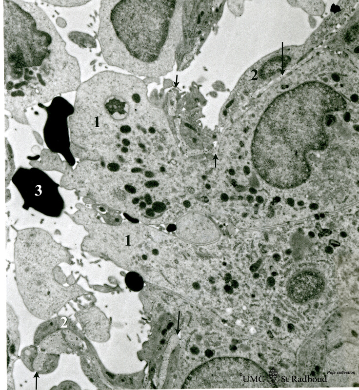
Title: Border of a marginal zone in spleen (rat)
Description: Stain: Immunoelectron microscopy (gold labelling of heparan sulfate in Lawyerly embedding, using the single chain antibody HS4C3).
The zone of red pulp immediately surrounding a lymphatic nodule is called the marginal zone (perilymphoid zone) and is composed of a scaffold of basal lamina material where a dendritic cell cytoplasm (APC) (1) and its cell projections are protruding into a capillary with thin lining cells (2) belonging to the system of anastomosing vascular channels at the border between the white and red pulp. (3) Erythrocyte in the red pulp.
Arrows indicate parts of sectioned scaffold consisting of thin basal lamina material where electron-dense particles of heparan sulfate molecules are localized (only visible by enlargement of this image). The glycosaminoglycan heparan sulfate is generally integrated in these scaffolds of basal laminae and is secreted by the lining cells.
Keywords/Mesh: lymphatic tissue, spleen, marginal zone, heparan sulphate, HS4C3 antibody, dendritic cell, histology, electron microscopy, POJa collection

