13.1 POJA-L4541+4542+4543
Title: Pericardium, epicardium and endocardium of the heart
Description:
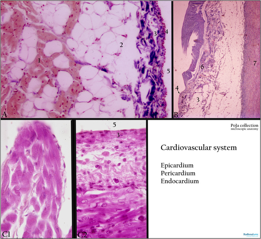
(A): Heart, epicardium, atrium, Azan, human. The myocardium (1) is enclosed by a subepicardial layer of adipose tissue (2) and a layer of connective tissue (3) covered by a thin layer of mesothelium (4) facing the lumen (5) of the pericardium. The atrial epicardium is thicker than that of the ventricle.
(B): Heart, part of the pericardium, Azan, human. The heart is a hollow muscular organ bathed in the pericardial fluid within a fibrous sac (pericardium). The pericardium is a mesothelial fold composed of regular dense connective tissue and lined by a single lining of mesothelial cells responsible for the production of pericardium fluid or liquor pericardii (up to 20 ml). Facing the epicardium is the tunica serosa or serous lamina (6) that also shows a thin mesothelium (4 arrow) followed by loose connective tissue mixed with adipose tissue (3). The outer layer is the tunica fibrosa or fibrous lamina (7) of dense regular connective tissue.
(C1-2): Heart, endocardium, Azan, human. Heart is a hollow organ with a lumen lined by a single endothelial cell layer and some connective tissue the so-called endocardium (3). A network of branched muscle cells in the connective tissue actively regulates the resistance due to diastolic processes. The endocardium is oxygen-supplied by the subendocardial capillaries, however in the left atrium/ventricle additional supply is provided by the oxygen-rich blood flow. In the areas of entrance and outflow of large blood vessels the endocardium is thick indicating that within the atria it is better developed than elsewhere. (C1) Thin endocardium (papillary muscle in ventricle); (C2) Thick endocardium (atrium). (5) Lumen of heart.
Background: Lymph vessels and both unmyelinated and myelinated nerve fibres run in the epicardium, myocardium and endocardium and especially in the subepicardial and subendocardial areas. Focally clusters of ganglion cells are present. During depolarisation the stimulus is carried from the endocardium to epicardium and vice versa during repolarisation. The cardiac endothelium comprises the endothelial cells of capillaries and that of the endocardium. This specialised endothelium has a modulatory role in the control of myocardial function because contractility and electrophysiological environment of the cardiomyocytes are regulated by this cardiac endothelium. It is also known that the endothelium controls embryonal heart development and is also implicated during hypertrophy in the adult. It is assumed that this cardiac endothelium may also act as a kind of Blood-Heart Barrier (analogous to the Blood-Brain-Barrier) as they control the ionic composition of the extracellular fluid around the cardiomyocytes (paracrine regulation), (D.L. Brutsaert Physiol Rev 2003, J. Noireaud and R. Andriantsitohaina BioMed Research International 2014. http://dx.doi.org/10.1155/2014/923805). http://www.hindawi.com/journals/bmri/2014/923805/
Keywords/Mesh: cardiovascular system, heart, ventricle, epicardium, pericardium, endocardium, mesothelium, cardiac endothelium, histology, POJA collection

