
5.4.3 POJA-L2374+2432+
2450+2447+2448+2444+
2445.
Collecting tubules (XVI) in the human kidney
5.4.3 POJA-L2374+2432+2450+2447+2448+2444+2445
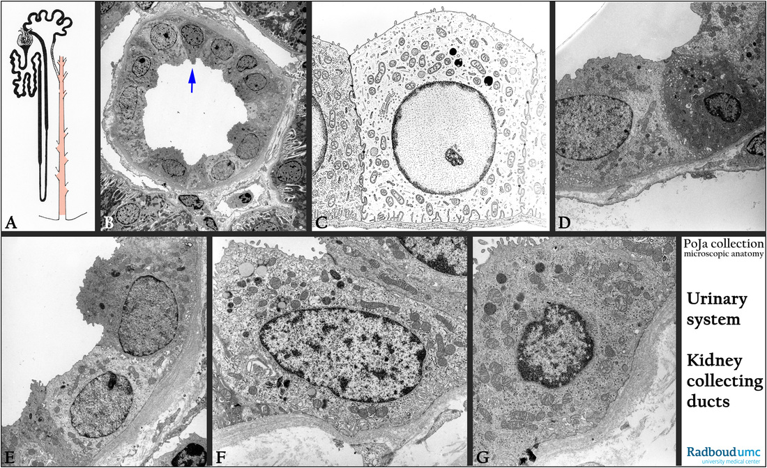
Title: Collecting tubules (XVI) in the human kidney
Description:
(A): Scheme of nephron. The collecting duct in the cortex is painted red-orange.
(B): Electron micrograph of a cortical collecting duct (CCD). The arrow points to a dark intercalated cell (IC).
(C): Electron microscopy scheme of a collecting duct cell (light principal cell), showing a limited number of mitochondria, basal invaginations and apical microvilli. At its left part of a dark principal cell. The basal lamina is often thick and laminated in human.
(D): Electron micrograph showing a dark intercalated cell (IC) with numerous apical vesicles. Adjacent a light principal cell.
(E): Electron microscopy, dark and light principal cells in a medullary ray. Note thick basal lamina.
(F): Electron micrograph of a light principal cell.
(G): Electron micrograph of a dark principal cell. The collecting tubules and ducts progressively concentrate the dilute urine by osmotic transfer of water into the hypertonic medullary interstitial tissue and subsequently towards the vasa recta.
Keywords/Mesh: urinary system, kidney, collecting duct, principal cell, intercalated cell, light principal cell, dark principal cell, histology, electron microscopy, POJA collection