
7.1 POJA-L1335+1741. Electron microscopy of the zona pellucida (gerbil, rabbit)
7.1 POJA-L1335+1741
Title: Electron microscopy of the zona pellucida (gerbil, rabbit)
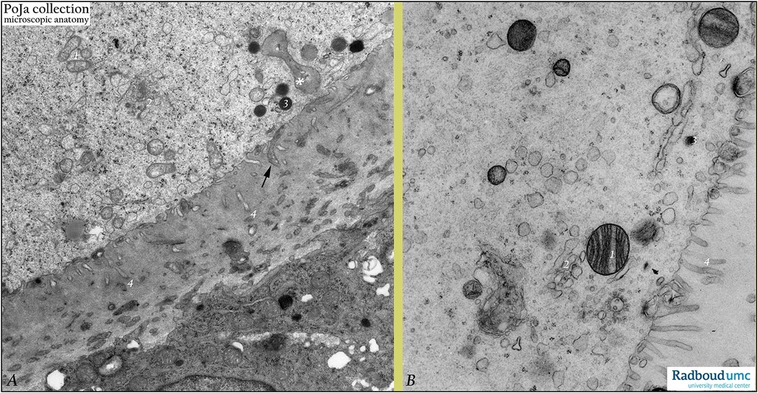
Description: Electron microscopy (A, gerbil; B, rabbit).
(A): Peripheral cytoplasm of oocyte of a secondary follicle (type 3). Few mitochondria (1), Golgi complexes (2) and electron-dense cortical granules (3), at (*) invagination of zona pellucida. The zona pellucida appears as a thick electron-grey basal lamina-like band (glycoproteins). Within this zone (4) slender processes of oocyte (electron-light) and granulosa cells are intermingled. Note long electron-darker apical villi (↑) of granulosa cell in close contact with oocyte surface.
(B): Peripheral cytoplasm of oocyte of a secondary follicle (type 4) with fewer organelles (1, 2, 3). Short microvilli of oolemma into zona pellucida (4).
Background: It is assumed that communication between oocyte and granulosa cells is bidirectional. The interplay is essential for development of oocyte as well as for follicle (granulosa) cells. This so-called oocyte-granulosa cell regulatory loop is important for differentiation of normal follicles as well as for the production of an oocyte that will be competent to undergo fertilization and embryogenesis. The oocyte is the dominant factor in establishment and maintenance of the cumulus lineage of granulosa cells. This oocyte-cumulus interaction prevents a.o. luteinization of cumulus cells by promoting growth, regulating steroidogenesis and inhibin synthesis, and suppressing LH receptor expression. The zona pellucida (ZP) plays an important role in this fertilization process; it is composed of glycoproteins ZPGp1- ZPGp2- ZPGp3- ZPGp4. Human ZP is probably similar in structure to murine ZP; it consists of long heterodimeric filaments of ZPGp2 and ZPGp3 which are cross-linked by homodimers of ZPGp. Human ZPGp1 consists of dimers of 100 kD. The ZPGp2 (75 kD), ZPGp3 (55 kD) and ZPGp4 (65 kD) form long filament complexes interconnected by ZPGp1 dimers in a regular way; ZPGp3, however, is involved in the binding process of spermatozoa. The ZP is formed by secretions from both oocyte and granulosa cells. The ZP contains four Gp’s (ZPGp’s 1 to 4) in human. They are synthesized, secreted and assembled during folliculogenesis. These ZPGp’s are assembled in a three-dimensional highly ordered filamentous structure. The result is a specialized extracellular multilaminar matrix surrounding the developing oocyte. It is known that capacitation (occurring while sperm reside in the female reproductive tract 1 up to 6 hours) includes the process of loss of spermatozoal glycocalyx as well as vesiculation of the acrosome of spermatozoa. During sperm binding oligosaccharides linked to the glycoprotein ZPGp3 (55 kD) of the zona pellucida (ZP) interact with sperm receptors. It appears that only acrosome-reacted spermatozoon can interact with ZPGp3. The cortical granules from the Golgi areas migrate to the periphery of the oocyte fuse with the oocyte plasma membrane (oolemma). The cortical reaction refers to the exocytosis of these granules discharging their contents (mixtures of proteases) in the ZP shortly after sperm-oocyte fusion. Proteases alter the pellucidal structure resulting in a specific transformation of the ZP that penetration by other capacitated spermatozoa is prevented (so-called zona reaction).
Keywords/Mesh: female reproductive organs, ovary, ovarian follicle, oocyte, zona pellucida, granulosa cells, female genitalia, electron microscopy, histology, POJA collection
Title: Electron microscopy of the zona pellucida (gerbil, rabbit)
Description: Electron microscopy (A, gerbil; B, rabbit).
(A): Peripheral cytoplasm of oocyte of a secondary follicle (type 3). Few mitochondria (1), Golgi complexes (2) and electron-dense cortical granules (3), at (*) invagination of zona pellucida. The zona pellucida appears as a thick electron-grey basal lamina-like band (glycoproteins). Within this zone (4) slender processes of oocyte (electron-light) and granulosa cells are intermingled. Note long electron-darker apical villi (↑) of granulosa cell in close contact with oocyte surface.
(B): Peripheral cytoplasm of oocyte of a secondary follicle (type 4) with fewer organelles (1, 2, 3). Short microvilli of oolemma into zona pellucida (4).
Background: It is assumed that communication between oocyte and granulosa cells is bidirectional. The interplay is essential for development of oocyte as well as for follicle (granulosa) cells. This so-called oocyte-granulosa cell regulatory loop is important for differentiation of normal follicles as well as for the production of an oocyte that will be competent to undergo fertilization and embryogenesis. The oocyte is the dominant factor in establishment and maintenance of the cumulus lineage of granulosa cells. This oocyte-cumulus interaction prevents a.o. luteinization of cumulus cells by promoting growth, regulating steroidogenesis and inhibin synthesis, and suppressing LH receptor expression. The zona pellucida (ZP) plays an important role in this fertilization process; it is composed of glycoproteins ZPGp1- ZPGp2- ZPGp3- ZPGp4. Human ZP is probably similar in structure to murine ZP; it consists of long heterodimeric filaments of ZPGp2 and ZPGp3 which are cross-linked by homodimers of ZPGp. Human ZPGp1 consists of dimers of 100 kD. The ZPGp2 (75 kD), ZPGp3 (55 kD) and ZPGp4 (65 kD) form long filament complexes interconnected by ZPGp1 dimers in a regular way; ZPGp3, however, is involved in the binding process of spermatozoa. The ZP is formed by secretions from both oocyte and granulosa cells. The ZP contains four Gp’s (ZPGp’s 1 to 4) in human. They are synthesized, secreted and assembled during folliculogenesis. These ZPGp’s are assembled in a three-dimensional highly ordered filamentous structure. The result is a specialized extracellular multilaminar matrix surrounding the developing oocyte. It is known that capacitation (occurring while sperm reside in the female reproductive tract 1 up to 6 hours) includes the process of loss of spermatozoal glycocalyx as well as vesiculation of the acrosome of spermatozoa. During sperm binding oligosaccharides linked to the glycoprotein ZPGp3 (55 kD) of the zona pellucida (ZP) interact with sperm receptors. It appears that only acrosome-reacted spermatozoon can interact with ZPGp3. The cortical granules from the Golgi areas migrate to the periphery of the oocyte fuse with the oocyte plasma membrane (oolemma). The cortical reaction refers to the exocytosis of these granules discharging their contents (mixtures of proteases) in the ZP shortly after sperm-oocyte fusion. Proteases alter the pellucidal structure resulting in a specific transformation of the ZP that penetration by other capacitated spermatozoa is prevented (so-called zona reaction).
Keywords/Mesh: female reproductive organs, ovary, ovarian follicle, oocyte, zona pellucida, granulosa cells, female genitalia, electron microscopy, histology, POJA collection