
14.1 POJA-L6247+6264+6242 Electron micrographs of cell organelles in striated muscle cells
14.1 POJA-L6247+6264+6242 Cell organelles in striated muscle cells
14.1 POJA-L6247+6264+6242 Electron micrographs of cell organelles in striated muscle cells
(B, C by courtesy of L. Eshuis BSc Section Electron microscopy, Department of Pathology, Radboud university medical center, Nijmegen, The Netherlands)
Title: Electron micrographs of cell organelles in striated muscle cells
Description:
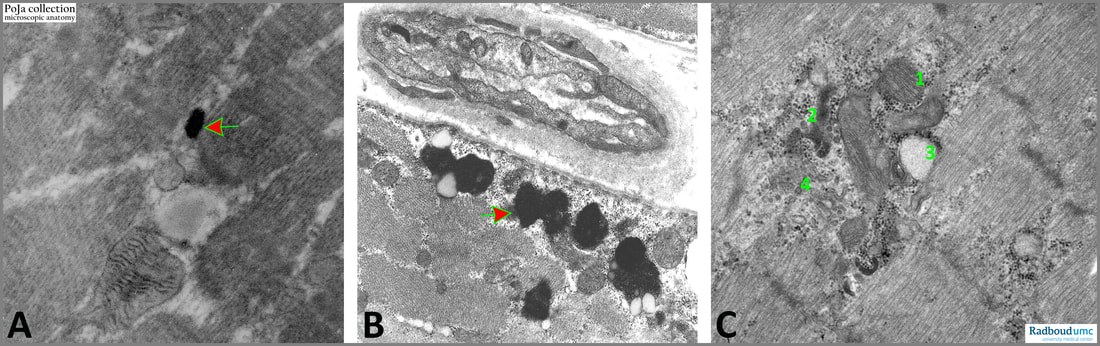
(A): Arrow indicates a small peroxisome in a diaminobenzidine stained section (rat). Peroxisomes are membrane-bound organelles (previously known as microbodies) and they are sparsely demonstrated in normal and healthy mammalian muscle. These organelles can be formed de novo from the endoplasmic reticulum and are involved in lipid catabolism in mammals. In humans, peroxisomes are responsible for the oxidation of specific classes of fatty acids e.g., the very-long-chain fatty acids that cannot be metabolized directly by mitochondria. They are also incorporated in the synthesis of bile acids and plasmalogens, detoxification of glyoxylate, and H2O2 metabolism. In this way skeletal muscle is responsible for lipid uptake and utilization, and it contributes significantly to whole-body lipid homeostasis.
(B): Lipofuscin granules peripherally in the sarcoplasm (arrow). In light microscopy they appear as yellow-brown granules (so-called wear and tear pigment, age pigment). Note the characteristic ultrastructural aspect of this residual body (lysosomal-related structure), it is a pleiomorphic membrane-bound body with an electron-dense granular composition and few lipid inclusions.
In the interstitium a capillary with pericyte is present, human.
(C): Golgi centrally in sarcoplasm (4); A dilated cistern (3); Small lysosomes (2); Mitochondria (1), human.
See also
Keywords/Mesh: locomotor system, skeletal muscle, striated muscle, peroxisome, lipofuscin, Golgi apparatus, electron microscopy, POJA collection
(B, C by courtesy of L. Eshuis BSc Section Electron microscopy, Department of Pathology, Radboud university medical center, Nijmegen, The Netherlands)
Title: Electron micrographs of cell organelles in striated muscle cells
Description:
(A): Arrow indicates a small peroxisome in a diaminobenzidine stained section (rat). Peroxisomes are membrane-bound organelles (previously known as microbodies) and they are sparsely demonstrated in normal and healthy mammalian muscle. These organelles can be formed de novo from the endoplasmic reticulum and are involved in lipid catabolism in mammals. In humans, peroxisomes are responsible for the oxidation of specific classes of fatty acids e.g., the very-long-chain fatty acids that cannot be metabolized directly by mitochondria. They are also incorporated in the synthesis of bile acids and plasmalogens, detoxification of glyoxylate, and H2O2 metabolism. In this way skeletal muscle is responsible for lipid uptake and utilization, and it contributes significantly to whole-body lipid homeostasis.
(B): Lipofuscin granules peripherally in the sarcoplasm (arrow). In light microscopy they appear as yellow-brown granules (so-called wear and tear pigment, age pigment). Note the characteristic ultrastructural aspect of this residual body (lysosomal-related structure), it is a pleiomorphic membrane-bound body with an electron-dense granular composition and few lipid inclusions.
In the interstitium a capillary with pericyte is present, human.
(C): Golgi centrally in sarcoplasm (4); A dilated cistern (3); Small lysosomes (2); Mitochondria (1), human.
See also
- 14.1 POJA-L6316+6317+6246 Autophagic vacuoles in striated muscle
Keywords/Mesh: locomotor system, skeletal muscle, striated muscle, peroxisome, lipofuscin, Golgi apparatus, electron microscopy, POJA collection