5.4.3 POJA-L2314+-2432+2373+4260+2459+2460
Title: Distal tubules (mTAL and cTAL) (VII) in kidney
Description:
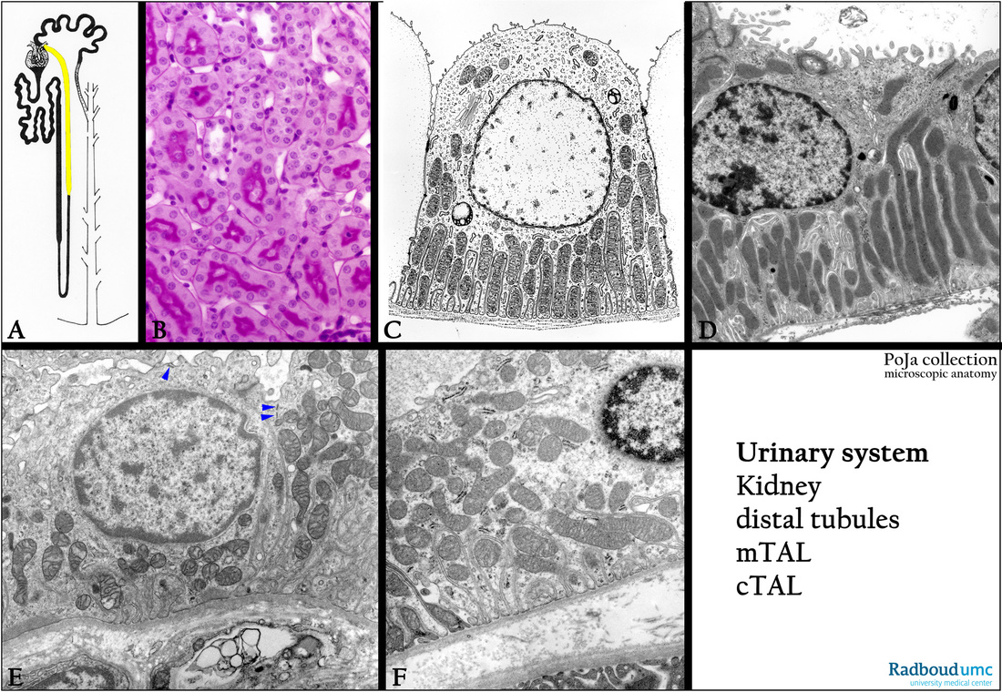
(A): Nephron, scheme, human. Yellow painted mTAL and cTAL parts of the distal tubules.
(B): Kidney cortex, stain PAS, human. PAS positive staining along the microvilli of the proximal convoluted tubule (PCT).
Distal tubules (cTAL) and cortical collecting ducts (CCD) are negative.
(C, D): Electron microscopy scheme of cell of a distal tubule, human. The cell lacks the numerous long microvilli as shown
in the proximal area. Numerous long mitochondria are arranged between the foldings of the basal lamina.
The electron micrograph of this cell type in rat kidney is shown in (D).
(E, F): Electron microscopy, human, the mitochondria in the cells of the cortical straight part of the thick ascending limb (cTAL) display
a less well arrangement between the folds of the basal labyrinth. Note also the apical tubulovesicular structures (arrowheads in E).
Keywords/Mesh: urinary system, kidney, distal tubule, cTAL, mTAL, histology, electron microscopy, POJA collection

