
6.2 POJA-L4240+4239+2714
+2715.
Testis and epididymides
6.2 POJA-L4240+4239+2714+2715
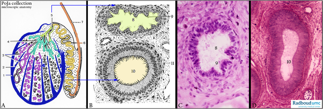
Title: Testis and epididymis
Description:
(A) Scheme testis with epididymis.
(B) Scheme efferent ductule and epidydimal duct.
(C) Stain hematoxylin-eosin, efferent ductule, human.
(D) Stain Weigert hematoxylin, epidydimal duct, human.
(A): The testis is packed within a dense connective tissue layer, the tunica albuginea (1), that further divides the testis in lobules
via septa (septulum testis) (2). (3) Lobuli testes with the seminiferous tubules. (4) Rete testis.
(5) Efferent ductules (ductuli efferentes), shown in detail in (B, 8), and in (C) where the scalloped outlined pseudostratified
epithelium and its irregular height are shown. Some of the columnar principal cells at the irregular protrusions bear stereocilia,
ciliated cells show kinocilia and microvilli (B, 9), interspersed a few basal cells.
(A, 6 and B, 10 and D, 10): Epidydimal duct (ductus epididymis). The epididymal duct is lined by two-layered pseudostratified
epithelium covered with stereocilia that look cluttered together sometimes (B, 11).
Smooth muscle fibers surround both the epididymal duct as well as the efferent ductules, needed for the expulsion of the ejaculate.
Keywords/Mesh: testis, epididymis, efferent ductule, epididymal duct, histology, POJA collection