
13.1 POJA-La0296+L4686+4698+4699
Arteries: physiological degeneration /inflammation (human)
13.1 POJA-La0296+L4686+4698+4699
Title: Arteries: physiological degeneration /inflammation (human)
Description:
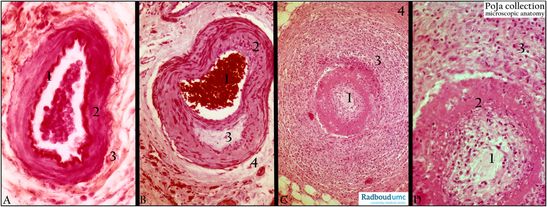
(A): Small muscular artery, omentum, human, (orcein-azocarmine staining). (1) Well-developed internal elastic membrane. (2) Fine branching and distribution of thin elastic fibers in media. (3) Adventitia with discontinuous external elastic membrane and few elastic fibres.
(B): Ring artery, uterus, myometrium, gravida, human, Azan staining. (1) Lumen of spiral artery with erythrocytes. (2) Media. (3) Hyalinisation in media. (4) Adventitial hyalinisation. Following gestation spiral arteries (branches of the uterine artery) show hyalinisation in the media and adventitia as signs of physiological degeneration.
(C, D): Vasculitis (Polyarteritis nodosa), kidney, human. Inflammation of blood vessel (kidney), Haematoxylin-eosin, human. Vasculitis of a mid-sized artery showing in (1) Lumen with edematous intima. (2) Fibrinoid necrosis. (3) Media with inflammatory cells. (4) Normal smooth muscle cells of the media.
Background: Polyarteritis nodosa (PAN) is an inflammation of small/mid-sized arteriae (vasculitis) that affects multiple organs as kidney, skin, gut, heart. The age of onset ranges from childhood to late adulthood. The disease has been associated with hepatitis B and/or C and therefore more common in injection drug addicts. PAN is probably mediated by deposition of immune complexes with viral antigens in blood and is also found deposited in inflamed vessels. Small aneurysms thread beads and give rise to the so-called ‘rosary sign’. In case of renal involvement eventually hypertension and kidney failure develops and rupture of renal microaneurysms occurs. In the first stages vasculitis of mostly arterioles is characterised by fibrinoid necrosis and leukocyte infiltration of the intima and media. The second stage is characterised by reabsorption of the fibrinoid and replacement by granulation tissue. Eventually in the third stage, granulation tissue develops into scar tissue with a narrowing of the lumen of the blood vessel. The inflammatory components and the scarring process may lead to obstruction in the circulation and even infarction (e.g. kidney, spleen infarctions).
Keywords/Mesh: cardiovascular system, vascularisation, blood vessel, arteriole , muscular artery, vasculitis, fibrinoid necrosis, inflammation, hyalinisation, periarteritis nodosa, histology, POJA collection
Title: Arteries: physiological degeneration /inflammation (human)
Description:
(A): Small muscular artery, omentum, human, (orcein-azocarmine staining). (1) Well-developed internal elastic membrane. (2) Fine branching and distribution of thin elastic fibers in media. (3) Adventitia with discontinuous external elastic membrane and few elastic fibres.
(B): Ring artery, uterus, myometrium, gravida, human, Azan staining. (1) Lumen of spiral artery with erythrocytes. (2) Media. (3) Hyalinisation in media. (4) Adventitial hyalinisation. Following gestation spiral arteries (branches of the uterine artery) show hyalinisation in the media and adventitia as signs of physiological degeneration.
(C, D): Vasculitis (Polyarteritis nodosa), kidney, human. Inflammation of blood vessel (kidney), Haematoxylin-eosin, human. Vasculitis of a mid-sized artery showing in (1) Lumen with edematous intima. (2) Fibrinoid necrosis. (3) Media with inflammatory cells. (4) Normal smooth muscle cells of the media.
Background: Polyarteritis nodosa (PAN) is an inflammation of small/mid-sized arteriae (vasculitis) that affects multiple organs as kidney, skin, gut, heart. The age of onset ranges from childhood to late adulthood. The disease has been associated with hepatitis B and/or C and therefore more common in injection drug addicts. PAN is probably mediated by deposition of immune complexes with viral antigens in blood and is also found deposited in inflamed vessels. Small aneurysms thread beads and give rise to the so-called ‘rosary sign’. In case of renal involvement eventually hypertension and kidney failure develops and rupture of renal microaneurysms occurs. In the first stages vasculitis of mostly arterioles is characterised by fibrinoid necrosis and leukocyte infiltration of the intima and media. The second stage is characterised by reabsorption of the fibrinoid and replacement by granulation tissue. Eventually in the third stage, granulation tissue develops into scar tissue with a narrowing of the lumen of the blood vessel. The inflammatory components and the scarring process may lead to obstruction in the circulation and even infarction (e.g. kidney, spleen infarctions).
Keywords/Mesh: cardiovascular system, vascularisation, blood vessel, arteriole , muscular artery, vasculitis, fibrinoid necrosis, inflammation, hyalinisation, periarteritis nodosa, histology, POJA collection