2.1 POJA-L932
Title: The effect of cyclophosphamide on the CD8-thymocytes in thymus (rat)
Description: Stain: Immunoperoxidase DAB.
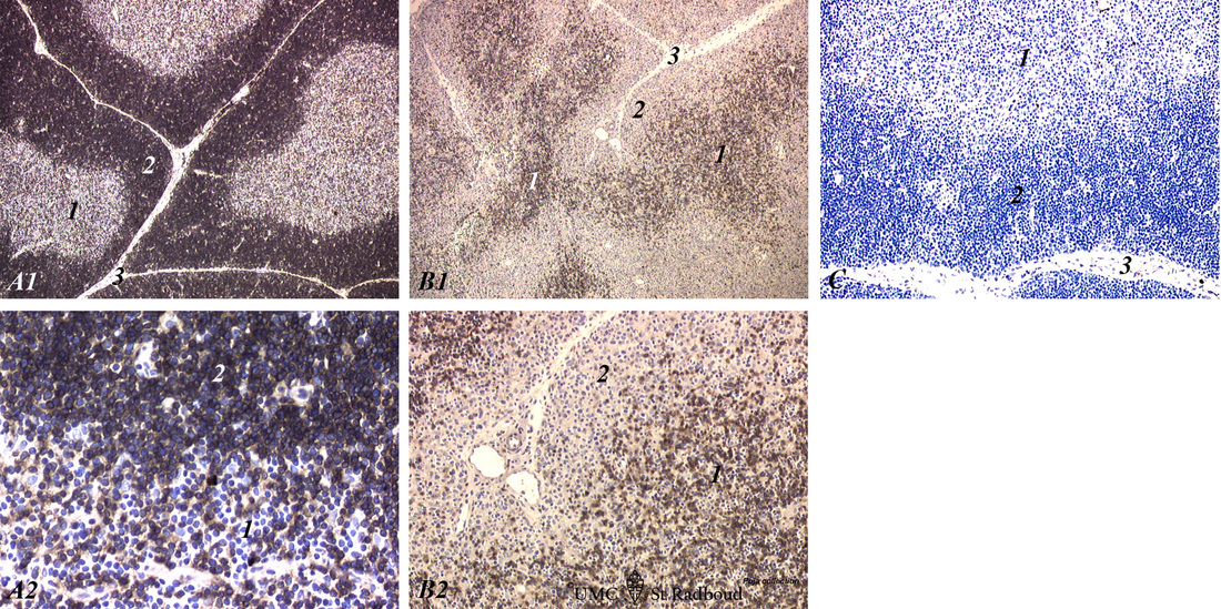
A single injection with cyclophosphamide (CP, 70 mg/ml) induces a transient cortical involution after 4 days, i.e. the darkly stained cortex and the lightly stained medulla in normal thymus (A1, A2) turn to darkly stained medulla and a lightly stained cortex (B1, B2) four days after a single injection with CP. The thymus recovers to normal about 10 days later.
(C) shows a negative Immunoperoxidase control of a normal thymus.
The anti CD8 monoclonal antibody in normal thymus tissue (A1, A2) shows an intense cortical staining, while in the medulla (A2-1) the positively stained cells are much less, since the medulla contains single-positive CD8 cells, while the cortex contains both double-positive (CD4+CD8+) and single positive lymphocytes (CDE8+).
The large CD4+ CD8+ population harbours the majority of dividing thymocytes.
Due to the CP-induced inhibition of cell proliferation in the cortex (B1, 2) the remaining cortical thymoblasts hardly express CD8, while the medullar thymocytes are strongly positive, con firming the so-called transient thymic inversion.
(A1, A2): Normal thymus;
(B1, B2): CP treated thymus;
(C): A negative Immunoperoxidase control.
(1): Medulla. (2): Cortex. (3): Septae or trabeculae.
Keywords/Mesh: lymphatic organs, thymus, CD8, cyclophosphamide, rat, toxicology, histology, POJA collection

