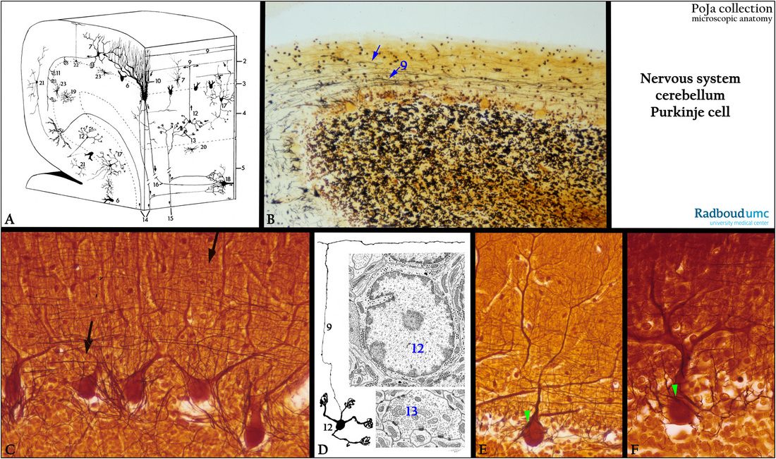
11.4 POJA-L4451+3105+3110+2973+3102+3103
Title: Cerebellum, Purkinje cells and granule cells
Description:
(A): Scheme cerebellum, human (zoom in).
Referred to Section 11.1 .
(1) Pia mater.
(2) Molecular layer.
(3) Ganglion layer with Purkinje cells.
(4) Granular (or granule) layer.
(5) Medulla rich in myelinated fibers (white matter).
(6) Purkinje cells.
(7) Basket cell (interneuron).
(8) absent in this scheme
(9) Parallel fibers (axons of granule cells).
(10) Dendrites of Purkinje cells in contact with climbing fibers.
(11) Fibers of basket cell around Purkinje cell.
(12) Granule cell (interneuron).
(13) Glomerulus (cerebellar island), contact place between mossy fibers and granule cells.
(14) Climbing fibers (input) ending on the proximal dendrites of Purkinje cells.
(15) Mossy fibers (input) ending in glomerulus.
(16) Axon of Purkinje cell (output from the cerebellar cortex).
(17) Golgi cell (interneuron).
(18) Neuron in deep cerebellar nucleus.
(19) Astrocyte in granular layer.
(20) Fibrous astrocyte in medulla.
(21) Outer stellate cell.
(22) Bergmann glial cell.
(23) Lugaro cell.
(B): Silver stain, human, transversal section. T-shaped axons (neurites) of the granule cells (arrows) form parallel fibers (9).
(C): Silver stain, human, sagittal section. Purkinje cells and T-shaped axons (parallel fibers) of granule cells (arrows).
(D): Electron microscopy scheme (human) of a granule cell (12), parallel fibers (9) and cerebral glomerulus (13) formed by axodendritic synapses of Golgi cell axons with granule cell dendrites. The granule cell (or granule neuron) is a nerve cell with a small soma and short dendrites with claw-shaped endings. The axon ascends into the molecular layer where it bifurcates and forms parallel fibers.
These axons run transversal (parallel to long axis of folium).
(E, F): Silver stain, human, sagittal section. Purkinje cell with dendrites and parallel fibers. Perpendicular to the parallel fibers basket cell axon and its descending collaterals run and form a pericellular basket (or brush or the ‘pinceau’) (green arrowheads) around the Purkinje cell body.
Keywords/Mesh: nervous tissue, cerebellum, gray matter, white matter, Purkinje cell, granule cell, basket cell, stellate cell, Golgi cell, parallel fiber, glomerulus, cerebellar island, GABA, histology, electron microscopy, POJA collection

