
14.1 POJA-L6086+6084 Ultrastructure of foetal muscle development (human)
14.1 POJA-L6086+6084 Foetal muscle development I
14.1 POJA-L6086+6084 Ultrastructure of foetal muscle development (human)
Title: Ultrastructure of foetal muscle development (human)
Description:
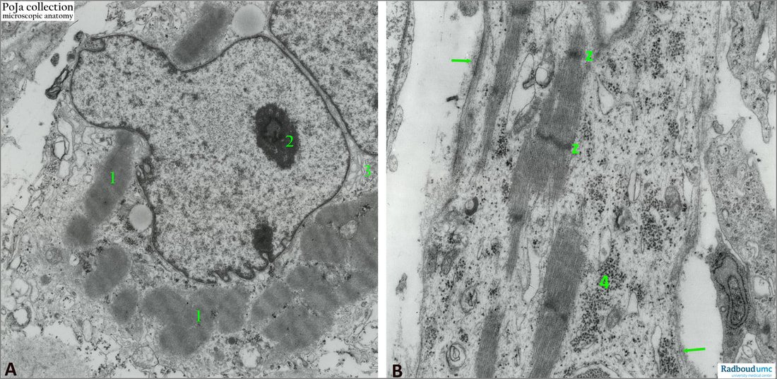
(A): Secondary myotube cell (12 weeks) showing condensed bundles of myofilaments (1). Lobulated nucleus with nucleolus (2). (3): Golgi area). Note small clusters of glycogen everywhere.
(B): Foetal myofilaments developing Z-lines. (4): Small areas of glycogen granules. Branches of growing myotubes are always present. (Arrows) point to thin flocculent, partly fibrous material at the outer side of the sarcoplasm as the formation of the future basal lamina.
Myoblast cell differentiation depends on myogenic determination factors such as MyoD, Mrf4, Myogenin and transcription factors as the MeF2 family.
Background:
Prenatal myogenesis is divided into primary and secondary myogenesis. During the embryonic stage and early foetal periods mainly primary myogenesis occurs with fusion of myoblasts into primary myotubes (in human during 3rd developmental week). These myotubes extend over the full length of the future muscle and attach to both tendons. As the muscle elongates, myoblasts will line up along the primary myotubes and eventually fuse to form secondary myotubes. Initially these tubes are formed between the ends of the primary myotubes but as they add new myoblasts there is a rapid growth in length. In human these secondary myotubes are formed from the 6th-8th developmental week. These fibres are multinucleated muscle cells that develops in close apposition to primary myotubes. Secondary myotubes use primary ones as template, they are formed on the surface of the primary ones beneath the basal lamina in the innervation zone of the muscle, and eventually attach to the muscle tendon. Both primary and secondary myotubes are innervated by the same motoneuron. Due to contraction secondary myotubes split away from the primary ones. The formation of myotubes is completed by the 18th week, after which most fibres are packed with myofibrils and show peripheral nuclei (Fidzianska 1971). The majority of adult muscle fibres is derived from secondary myotubes.
See also:
Keywords/Mesh: locomotor system, skeletal muscle, striated muscle, muscle development, myotube, electron microscopy, POJA collection
Description:
(A): Secondary myotube cell (12 weeks) showing condensed bundles of myofilaments (1). Lobulated nucleus with nucleolus (2). (3): Golgi area). Note small clusters of glycogen everywhere.
(B): Foetal myofilaments developing Z-lines. (4): Small areas of glycogen granules. Branches of growing myotubes are always present. (Arrows) point to thin flocculent, partly fibrous material at the outer side of the sarcoplasm as the formation of the future basal lamina.
Myoblast cell differentiation depends on myogenic determination factors such as MyoD, Mrf4, Myogenin and transcription factors as the MeF2 family.
Background:
Prenatal myogenesis is divided into primary and secondary myogenesis. During the embryonic stage and early foetal periods mainly primary myogenesis occurs with fusion of myoblasts into primary myotubes (in human during 3rd developmental week). These myotubes extend over the full length of the future muscle and attach to both tendons. As the muscle elongates, myoblasts will line up along the primary myotubes and eventually fuse to form secondary myotubes. Initially these tubes are formed between the ends of the primary myotubes but as they add new myoblasts there is a rapid growth in length. In human these secondary myotubes are formed from the 6th-8th developmental week. These fibres are multinucleated muscle cells that develops in close apposition to primary myotubes. Secondary myotubes use primary ones as template, they are formed on the surface of the primary ones beneath the basal lamina in the innervation zone of the muscle, and eventually attach to the muscle tendon. Both primary and secondary myotubes are innervated by the same motoneuron. Due to contraction secondary myotubes split away from the primary ones. The formation of myotubes is completed by the 18th week, after which most fibres are packed with myofibrils and show peripheral nuclei (Fidzianska 1971). The majority of adult muscle fibres is derived from secondary myotubes.
See also:
- Developmental programming of fetal skeletal muscle and adipose tissue development. Yan, X., Zhu, M-J., Dodson, M.V., Du, M. J Genomics 2013, 1, 29-38 https://www.jgenomics.com/v01p0029.htm
Keywords/Mesh: locomotor system, skeletal muscle, striated muscle, muscle development, myotube, electron microscopy, POJA collection