10.7 POJA-L2018+4366+4382+4371+4372+4381
Title: Melanoma skin
Description:
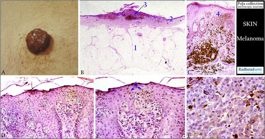
(A): Macroscopy skin of a nodular melanoma, human.
(B): Survey of nodular melanoma in the skin, a common superficial melanoma type. (B - F): Stain hematoylin-eosin, human.
(1) Subcutis, adipose cells. (2) Epidermis. (3) Melanoma.
(C): Abnormal junctional activity (4) of nevus cells, migrating upwards close to the epidermal cells.
“Junctional activity” is considered as the proliferation along the dermal-epidermal junction.
(D, E): The nevus cells have migrated very close to the dermal cells up to the stratum granulosum cell layer (junctional activity).
A detail comprise both nevus cells and melanoma cells (4), the single melanoma cells migrate upwards to the granular cell layer
of the skin.
(F): Detail of the melanoma cells. Melanoma cells are often large, epitheloid, polygonal or spindle shape with pleiomorphic nuclei.
Note the abundance and irregular pigmentation of the tumor cells.
(Micrographs (A, B) by courtesy of D. Ruiter, MD PhD, former Head Department of Pathology, Radboud university medical center, Nijmegen, The Netherlands)
Keywords/Mesh: skin, nodular melanoma, nevus cell, histology, pathology, macroscopy, POJA collection

