4.1.1 POJA-L4021+4017+La0330+4063
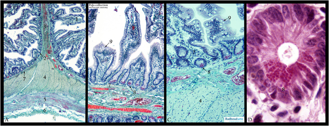
Title: Plica, villi and crypts in jejunum (human)
Description: Stain: (A, B, C) Trichrome Goldner. (D) Hematoxylin-eosin.
(A): Shows a Kerckring’s plica as a circular fold of mucosa and submucosa (3). The plica is studded with villi (1) and crypts (2), and supported by a loosely connective tissue layer, the lamina propria (7). The plica and intestinal tube is surrounded by a double muscle coat consisting of an inner circular muscle layer (4) and an outer muscle layer (5), followed by a tela subserosa (6) needed to embed the “tube” in its environment.
(B, C): Show details of the villi and crypts. On the bottom of the majority of the crypts Paneth cells are found having large characteristic refractile and intensely stained granules (8), detailed in (D).
The Paneth cells are known to produce α-defensins (or cryptdinsins), lysozymes, tumor necrosis factor and phospholipase A2, all involved in antimicrobial activity, thus contributing to a gastrointestinal defense barrier. It is thought that mucosal T-cells regulate the activity of the Paneth cells. (9) Points to the goblet cells present in the epithelial lining.
The fourth cell type, i.e. enteroendocrine cells, is not shown in these figures.
Keywords/Mesh: jejunum, plica, crypts, Paneth cells, goblet cells, enterocytes. histology, POJA collection

