
6.5 POJA-L4248B+4299+
4300+4301.
Penises
6.5 POJA-L4248B+4299+4300+4301
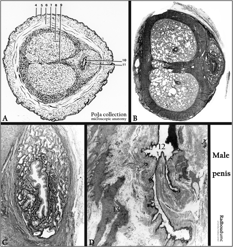
Title: Penis
Description:
(A) Scheme crossed-sectioned penis.
(B, C) Stain Masson, black-and-white micrograph of cross-sectioned penis with detail of urethra-seminal duct, human.
(D) Electron micrograph of area in corpus cavernosum, human.
The penis consists of two corpora cavernosa (8) and one corpus spongiosum (11) that surrounds the urethra-seminal duct (10), all ending up in the glans penis. All corpora are surrounded with a dense connective tissue sheaths, the tunica albuginea (7). A septum (9) divides the two corpora cavernosa. (6) The skin surrounds the tunica albuginea, and shows a multilayered, keratinizing squamous epithelium (4), followed by the cutis (5) and muscle cell fibers in the subcutis (6). The corpora in fact consist of labyrinth-like spaces (12) or cisterns lined with endothelium and formed by trabeculae of connective tissue and smooth muscle fibers (see also D, 13). These blood spaces are filled and drained by muscular helicine arteries (corpus cavernosum), urethral arteries (corpus spongiosum) and the accompanying venae.
Keywords/Mesh: penis, cavernous body, corpus spongiosum, corpus cavernosum, urethra, histology, electron microscopy, POJA collection