2.1 POJA-L924
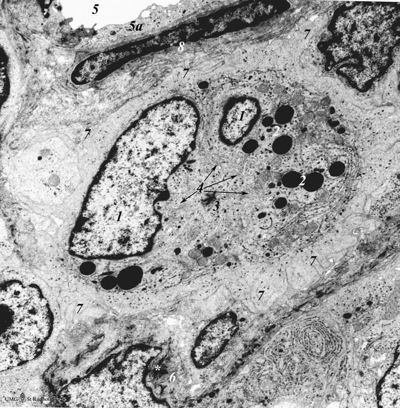
Title: Thymus medulla (rat, neonate)
Description: Electron microscopy.
An interdigitating cell in the thymic corticomedullary region shows a large electron-light cytoplasm with a complex branching (7) at the periphery.
(1): The nucleus is cut twice .
(2): There is abundance of organelles as well as of quite uniform electron-dense lysosomal structures.
(4): Note many small dark tubulovesicular structures.
(3): The region of the centrosome.
(5): Part of a blood vessel (5) with endothelium (5a) and an apposing pericyte (8).
(6): Medullar epithelioreticular cells with intermediate filaments (cytokeratin filaments,*).
Keywords/Mesh: lymphatic tissue, thymus, interdigitating cell, medullar epithelioreticular cell, histology, electron microscopy, POJA collection

