
10.3 POJA-L2223+2246+2261
+2229+2230+2262.
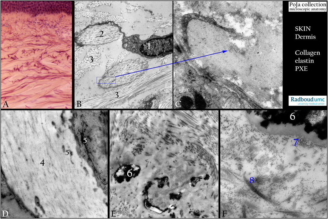
Collagen and elastin arrangement in the dermis of skin
10.3 POJA-L2223+2246+2261+2229+2230+2262
Title: Collagen and elastin arrangement in the dermis of skin
Description:
(A): Skin belly, stain resorcin-hematoxylin Weigert, human, visualizing mainly the elastine fibers, detailed in (B).
(B-D): Back skin, electron microscopy.
(B): Elastoblast (1) (fibroblast producing proelastin) with elastin fibrils (2) and striped aspect of denser-stained microfibrillar-
associated glycoproteins (MAGPs) and fibrillins, human. Collagen fibrils cross sectioned and longitudinal (3).
The blue rectangle in (B) is detailed in (C).
(C): Elastoblast with amorph whitish elastin fibril and peripheral localized MAGPs/fibrillins (arrow point ending) in the dermis
of rat.
(D): Large elastin fiber with faintly-stained internal localized MAGPs (4) as well as distinct denser stained peripheral fibrillary MAGPs/fibrillins (5), human.
(E-F): Electron microscopy. Abnormal elastic fiber in dermis of the back skin of a patient with pseudoxanthoma elasticum (PXE).
Brittle elastin is the result due to calcified elastin (6). They are surrounded by remnants of fibroblasts and slightly altered collagen fibrils.
At higher magnification the calcified zone (6), at (7) almost tubular-type MAPGs/fibrillins at the periphery.
(8)Sometimes abnormal frayed collagen fibers are present in the PXE-affected areas.
Keywords/Mesh: skin, dermis, fibroblast, elastoblast, collagen, elastin, fibrillin, glycoprotein, pseudoxanthoma elasticum, histology, electron microscopy, POJA collection