
13.1 POJA-L4319+4668 High endothelial venules (lymphoid organs)
13.1 POJA-L4319+4668
Title: High endothelial venules (lymphoid organs)
Description:
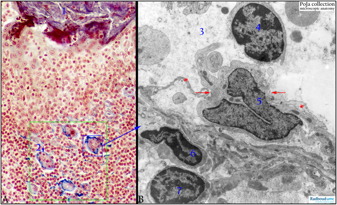
(A, B): Postcapillary venules (palatine tonsil, lymph node), Azan (human) resp. EM (dog).
(A): Reticular tissue with lymphocytes of the palatine tonsil. (1) Cellular debris with neutrophilic granulocytes. The green rectangle contains several HEVs (high endothelial venules) in contact with the sinusoids. The venule (2) shows several lymphocytes (intense red nucleus) (arrow) in diapedesis through the endothelial cells (large pale nucleus with nucleolus).
(Background: For more images see POJA Collection Microscopic Anatomy, Section 2.1 Lymphatic Organs)
(B): Venule in lymphatic nodule with lymphocyte in diapedesis. Diapedesis of a large lymphocyte (5) between the endothelial processes (**), indicated by red arrows. (3) Lumen of venule. (4) Small lymphocyte in lumen. (6, 7) Small lymphocytes outside the venule.
Background: Among the special types of blood vessels, the so-called high-endothelial venules (HEV) are known as regions where recirculating lymphocytes adhere (specifically) to the endothelial cells and subsequently migrate into the lymph node, a process called homing.
Keywords/Mesh: cardiovascular system, vascularisation, lymphatic tissue sinusoid, postcapillary venule, diapedesis, lymphocyte, histology, electron microscopy, POJA collection
Title: High endothelial venules (lymphoid organs)
Description:
(A, B): Postcapillary venules (palatine tonsil, lymph node), Azan (human) resp. EM (dog).
(A): Reticular tissue with lymphocytes of the palatine tonsil. (1) Cellular debris with neutrophilic granulocytes. The green rectangle contains several HEVs (high endothelial venules) in contact with the sinusoids. The venule (2) shows several lymphocytes (intense red nucleus) (arrow) in diapedesis through the endothelial cells (large pale nucleus with nucleolus).
(Background: For more images see POJA Collection Microscopic Anatomy, Section 2.1 Lymphatic Organs)
(B): Venule in lymphatic nodule with lymphocyte in diapedesis. Diapedesis of a large lymphocyte (5) between the endothelial processes (**), indicated by red arrows. (3) Lumen of venule. (4) Small lymphocyte in lumen. (6, 7) Small lymphocytes outside the venule.
Background: Among the special types of blood vessels, the so-called high-endothelial venules (HEV) are known as regions where recirculating lymphocytes adhere (specifically) to the endothelial cells and subsequently migrate into the lymph node, a process called homing.
Keywords/Mesh: cardiovascular system, vascularisation, lymphatic tissue sinusoid, postcapillary venule, diapedesis, lymphocyte, histology, electron microscopy, POJA collection