
7.1 POJA-L1930+1932+1940+1942+1658. Clear cell carcinoma of ovary (human, adult)
7.1 POJA-L1930+1932+1940+1942+1658
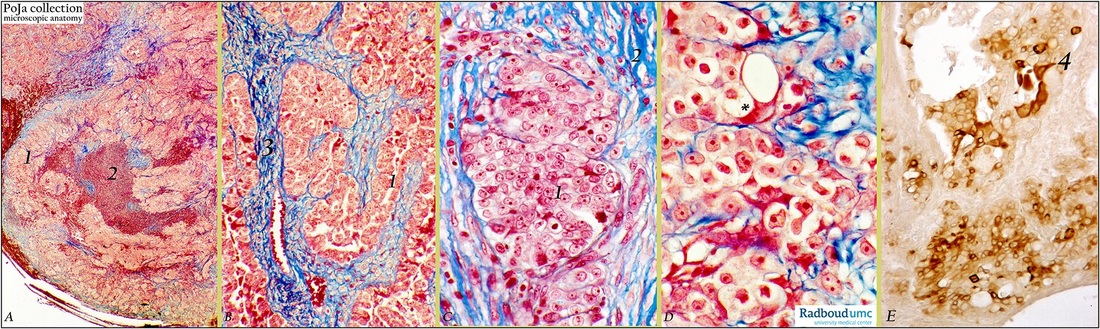
Title: Clear cell carcinoma of ovary (human, adult)
Description: (A, B. C, D) Stain: Mallory; (E) CK7 (OVTL12/30) antikeratin-7 antibody immunoperoxidase staining with diaminobenzidin reaction (DAB) and hematoxylin counterstaining.
(A): Low magnification show mixture of growth forms of neoplastic cells (1) and large hemorrhagic areas (2).
(B): Tubular formations (1) and blue-stained collagenous stroma and blood vessels (3).
(C): Nest of large light-stained epithelial neoplastic cells (1) and distinct nuclei; (2) stroma.
(D): Cytoplasms are clear (*) (presence of glycogen, that is washed out during fixation procedures). Cells show well demarcated cell borders with polymorphic nuclei.
(E): Heterogeneous staining with CK7 antibody. Note the characteristic hobnail cell(s) (4) that are stained conspicuously positive.
Background: Clear cell ovarian tumors are part of the surface epithelial-stromal tumor group of ovarian cancers. Sometimes presence of these tumors is found associated with endometriosis or endometrioid carcinoma of the ovary with a resemblance of the endometrial clear cell carcinomas. Embryologically the coelomic epithelium give rise to the müllerian epithelium (ducts of Müller) and its derivates are the fallopian tube lining (tubal pathway), endometrial lining (endometrial pathway) and endocervical glands lining (endocervical pathway). The ovarian surface epithelium contains undifferentiated cells that still are latent to differentiate along similar embryonic pathways. In this way a tumor derived from these cells will differentiate in similar way i.e. along the endometrial pathway. Therefore it is thought that these clear cell carcinomas are of müllerian duct origin and variants of endometrioid adenocarcinomas. These ovarian clear adenocarcinomas are relatively rare; most of them are malignant and account for 5-12% of all ovarian cancers. Usually they are large and firm with cysts and solid areas. A mixture of papillary, cystic, glandular or nest growth forms is found in these neoplasms. In the solid forms cells are arranged in sheets or tubules. The tumor is characterized by large epithelial cells with abundant clear cytoplasm (glycogen), sharply well-demarcated cell borders and hyperchromatic, pleiomorphic nuclei. Cysts and acini are lined by clear neoplastic cells; some of them protrude into the lumen (so-called hobnail cells with a nucleus standing on a stalk of cytoplasm) after secretion of glycogen.
Keywords/Mesh: female reproductive organs, ovary, clear cell carcinoma, female genitalia, ovarian neoplasms, adenocarcinoma, clear cell, keratin 7, OVTL12/30 antibody, histology, pathology, histology, POJA collection