
5.5. POJA-L2481+5005+2482.
Interstitial structure I of the cortical area of the kidney
5.5. POJA-L2481+5005+2482
Title: Interstitial structure I of the cortical area of the human kidney
Description:
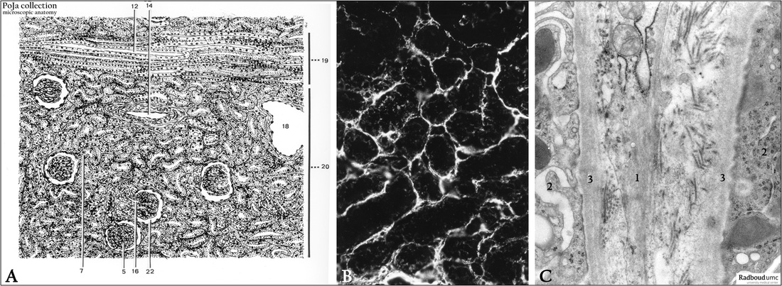
(A): Kidney cortex, scheme, human.
(5) Glomerulus.
(7) Urinary pole where the urinary space empties into the first part of the tubular system, pars contorta I, proximal tubule (PCT).
(12) Collecting tubule.
(14) Arteria corticalis radiata (cortical radial artery).
(16) Afferent arteriole.
(18) Vena arcuata (arcuate vein).
(19) Medulla radiata (medullary rays).
(20) Cortical labyrinth.
(22) Bowman’s space.
(B): Immunofluorescence with antibodies against smooth muscle actin showing their presence in the interstitial cell types between
the tubules, but not in the tubules.
(C): Electron microscopy. Between the two tubules (2) with their basal lamina (3) a peritubular myofibroblast (or interstitial cell, 1)
with actin filaments is highlighted.
Keywords/Mesh: urinary system, kidney, interstitium, myofibroblast, smooth muscle actin, histology, electron microscopy, POJA collection
Title: Interstitial structure I of the cortical area of the human kidney
Description:
(A): Kidney cortex, scheme, human.
(5) Glomerulus.
(7) Urinary pole where the urinary space empties into the first part of the tubular system, pars contorta I, proximal tubule (PCT).
(12) Collecting tubule.
(14) Arteria corticalis radiata (cortical radial artery).
(16) Afferent arteriole.
(18) Vena arcuata (arcuate vein).
(19) Medulla radiata (medullary rays).
(20) Cortical labyrinth.
(22) Bowman’s space.
(B): Immunofluorescence with antibodies against smooth muscle actin showing their presence in the interstitial cell types between
the tubules, but not in the tubules.
(C): Electron microscopy. Between the two tubules (2) with their basal lamina (3) a peritubular myofibroblast (or interstitial cell, 1)
with actin filaments is highlighted.
Keywords/Mesh: urinary system, kidney, interstitium, myofibroblast, smooth muscle actin, histology, electron microscopy, POJA collection