
5.4.1 POJA-L2944+2369+
2367+2366+2359+5035.
Mesangium cells in the renal glomerulus (XIV) of the kidney
5.4.1 POJA-L2944+2369+2367+2366+2359+5035
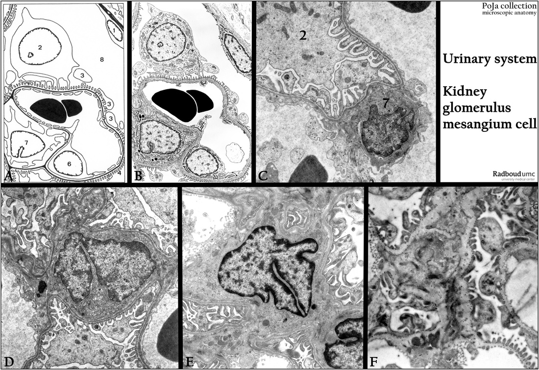
Title: Mesangium cells in the renal glomerulus (XIV) of the kidney
Description:
(A, B): Scheme of the glomerular podocyte and mesangium cell, human.
(1) Parietal cell of the Bowman capsule.
(2) Podocyte with pedicles (3, 4).
(5) Glomerular basement membrane (or lamina).
(6) Endothelial cell of capillary.
(7) Mesangium cell.
(8) Primary urine space.
(C): Electron microscopy with a podocyte (2) and a mesangium cell (7), rat.
(D, E): Electron microscopy showing centrally mesangium cells, rat.
(F): Eelctron microscopy, immunogold staining with antibodies (HS4C3) against heparan sulfate in the mesangial matrix and basal laminae, rat. The 10 nm gold labeling are found in the GBL of the capillaries, close to the pedicles as well as wide spread in the mesangium.
Background: The intraglomerular mesangium cells are of smooth muscle cell origin (vimentin, actin and desmin positive).
They can regulate the blood flow of the glomerular capillaries (contractile). They can also phagocytize immune complexes and
might be involved in monitoring blood-glucose levels. They form the mesangial matrix in which HSPG’s are found.
Keywords/Mesh: urinary system, kidney, glomerulus, glomerular basal lamina, podocyte, pedicle, mesangium cell, mesangium, heparan sulfate, histology, electron microscopy, POJA collection