
7.1 POJA-L1500+L1738+L1844
Corpora lutea of pregnancy, ovary (human)
7.1 POJA-L1500+L1738+L1844
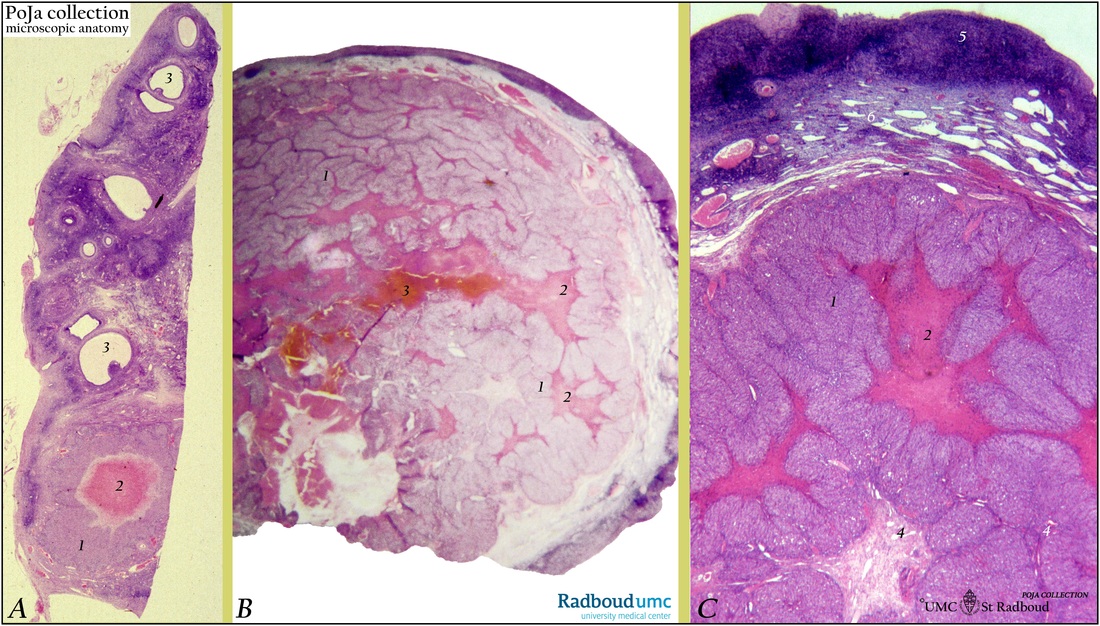
Title: Corpora lutea of pregnancy, ovary (human)
Description: (A, B, C) Stain: Hematoxylin-eosin.
(A): Survey of part of ovary, compare diameter of corpus luteum and antral follicles.
A large corpus luteum verum (of pregnancy) shows thick layer of epithelioid lutein cells (1) that surrounds the old follicular cavity, centrally core of fibrin (2) is present. Tertiary follicles i.e. antral follicles type 5 and 5+ at (3).
(B): Low magnification of part of corpus luteum verum in earlier stage. The corpus already shows characteristic loosely curling and folded bands of epithelioid walls (1) arranged around lakes of fibrin (2) and a more centrally located blood clot (3).
(C): Higher magnification of a more advanced stage of corpus luteum verum still shows coagulum composed of fibrin (2) filling the central cavity. The parenchymal band of lutein cells (1) is folded with ingrown fibrous septa richly supplied with thin-walled blood vessels (4). Cortex at (5) and medulla (6) of ovary.
Background: After ovulation the lining stratum granulosum (mural granulosa cells) of the empty follicle collapses and becomes folded. The result is a central fresh blood clot surrounded by loosely curling bands of lutein cells as the former granulosa cells transform and form epithelioid sheets. Under influence of luteinizing hormone (LH) the luteinization, process starts i.e. the granulosa cells increase in size and differentiate into granulosa lutein cells while they produce a yellow carotenoid pigment (lutein) as well as progesterone/estrogen (in response to follicle-stimulating-hormone FSH and LH stimulation). Hence the name corpus luteum due to the yellowish color and such a corpus luteum verum may reach up to 25 mm until the fourth month. Meanwhile the basement membrane of the collapsed follicle is broken down. Internal theca cells invade into the cellular mass together with capillaries/connective tissue and gradually old blood coagulum inside the former follicular cavity is replaced.
Keywords/Mesh: female reproductive organs, ovary, corpus luteum, female genitalia, pregnancy, antral follicles, luteinizing hormone, histology, POJA collection