7.2 POJA-L1538+1539+1866+1901
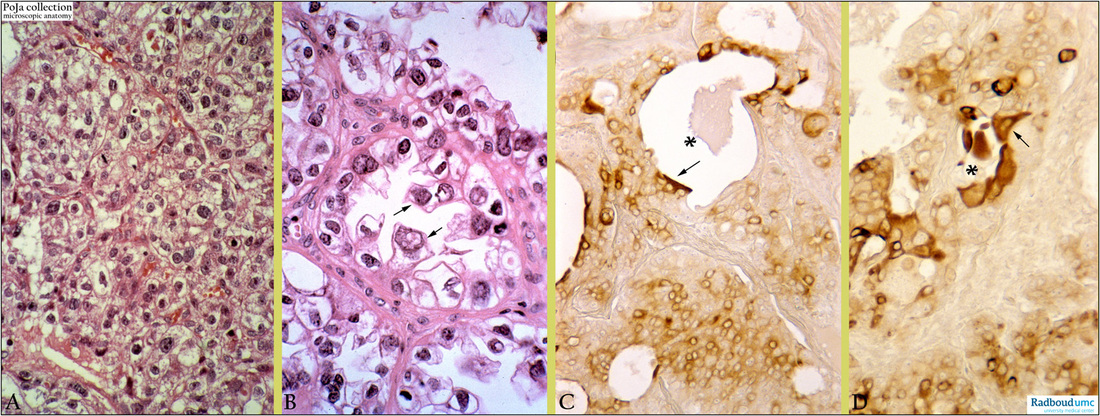
Title: Clear cell carcinoma, vagina (human, adult)
Description: Stain: (A-B) Hematoxylin-eosin; (C) CK 7 (OVTL 12-30) antikeratin antibody immunoperoxidase staining with diaminobenzidin reaction (DAB).
(A): Moderately polymorphic tumor cells with clear and vacuolized cytoplasm and well-defined cell borders are arranged in atypical glandular structures. Thin connective septa with blood vessels are localized between clusters of tumor cells.
(B): Moderate polyphormic tumor cells are polygonal up to columnar-shaped and their cytoplasm is clear due to glycogenation (so-called ‘clear’ cells). Nuclei with multiple irregular nucleoli are oval up to irregular shaped and frequently localized at the apex and are therefore called ‘hobnail’ cells being frequently observed (→).
(C-D): Tumor cells arranged in atypic acini (lumen, *) demonstrate heterogeneous staining for CK 7. Surprisingly ‘hobnail’ cells (→) are outstandingly positive for CK 7 expression. (Partly by courtesy of G. P. Vooijs MD PhD, former Head of the Department of Pathology, Radboud university medical center, Nijmegen, The Netherlands).
Keywords/Mesh: female reproductive organs, vagina, clear cell carcinoma, vaginal neoplasm, adenocarcinoma, clear cell carcinoma, cytokeratin 7, hobnail cells, histology, POJA collection

