
12.1.4 POJA-L4418+3569+
2572
Retina in the eye
12.1.4 POJA-L4418+3569+2572
Title: Retina I
Description:
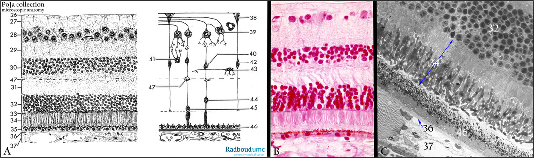
(A): Retina, schemes, human
(B): Retina, stain Azan, human.
(C): Retina, electron micrograph, gerbil. (A-C, compare identical layers)
(26) Inner limiting membrane (membrane limitans interna).
(27) Optic nerve fibre layer with anchoring parts of Müller cells (stratum neurofibrarum).
(28) Multipolar ganglion cell layer (3rd neuron) (stratum ganglionicum nervi optici).
(29) Inner plexiform layer (stratum plexiforme internum), i.e. the zone with synapses between bipolar
and multipolar ganglia cells.
(30) Stratum granulosum internum, or inner nuclear layer (2nd neuron) showing the nuclei of bipolar
ganglion cells, amacrine/horizontal/Müller cells.
(31) Outer plexiform layer ( stratum plexiforme externum) with synapses between bipolar ganglion cells,
photoreceptor cells and horizontal cells.
(32) Stratum granulosum externum (stratum neuroepitheliale or outer nuclear layer, 1st neuron).
(33) Membrana limitans externa or outer limiting membrane.
(34) Inner and outer segments of rods and cones.
(35) Pigmented epithelium or stratum pigmentosum retinae.
(36) Lamina basalis.
(37) Lamina choriocapillaris or choroidal capillary layer.
Compare both schemes (A):
(38 is 26) Basis (of Müller’s cell) on lamina basalis (inner limiting membrane).
(39 is 28) Multipolar ganglion cell.
(40 is 30) Bipolar ganglion cell.
(41 is 30) Amacrine cell.
(42 is 30) Müller cell.
(43 is 30) Horizontal cell.
(44 is 32) Rod cell.
(45 is 32) Cone cell.
(46 is 35) Pigmented epithelial cell.
(47 within 31) Synapses of cone cell as well as rod cell with bipolar ganglion cell.
Keywords/Mesh: eye, retina, choroid, rod, cone, histology, electron microscopy, POJA collection
Title: Retina I
Description:
(A): Retina, schemes, human
(B): Retina, stain Azan, human.
(C): Retina, electron micrograph, gerbil. (A-C, compare identical layers)
(26) Inner limiting membrane (membrane limitans interna).
(27) Optic nerve fibre layer with anchoring parts of Müller cells (stratum neurofibrarum).
(28) Multipolar ganglion cell layer (3rd neuron) (stratum ganglionicum nervi optici).
(29) Inner plexiform layer (stratum plexiforme internum), i.e. the zone with synapses between bipolar
and multipolar ganglia cells.
(30) Stratum granulosum internum, or inner nuclear layer (2nd neuron) showing the nuclei of bipolar
ganglion cells, amacrine/horizontal/Müller cells.
(31) Outer plexiform layer ( stratum plexiforme externum) with synapses between bipolar ganglion cells,
photoreceptor cells and horizontal cells.
(32) Stratum granulosum externum (stratum neuroepitheliale or outer nuclear layer, 1st neuron).
(33) Membrana limitans externa or outer limiting membrane.
(34) Inner and outer segments of rods and cones.
(35) Pigmented epithelium or stratum pigmentosum retinae.
(36) Lamina basalis.
(37) Lamina choriocapillaris or choroidal capillary layer.
Compare both schemes (A):
(38 is 26) Basis (of Müller’s cell) on lamina basalis (inner limiting membrane).
(39 is 28) Multipolar ganglion cell.
(40 is 30) Bipolar ganglion cell.
(41 is 30) Amacrine cell.
(42 is 30) Müller cell.
(43 is 30) Horizontal cell.
(44 is 32) Rod cell.
(45 is 32) Cone cell.
(46 is 35) Pigmented epithelial cell.
(47 within 31) Synapses of cone cell as well as rod cell with bipolar ganglion cell.
Keywords/Mesh: eye, retina, choroid, rod, cone, histology, electron microscopy, POJA collection