
12.1.2 POJA-L4393+2535+
4394+2537
Cornea and limbal area of the eye
12.1.2 POJA-L4393+2535+4394+2537
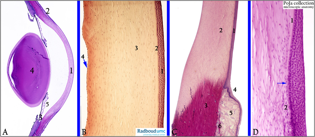
Title: Cornea and limbal area of the eye
Description:
(A): Survey, stain Haematoxylin-eosin, human. (1) Cornea. (2) Limbal area being the transition zone from the clear cornea into the opaque
scleral collagenous stroma. (3) Ciliary body with ciliary processes. (4) Lens. (5) Iris.
(B): The transparent cornea, stain Haematoxylin-azophloxine, human. The anterior corneal epithelium (1) is a non-keratinising squamous epithelium of 5 to 6 cell layers resting on the Bowman’s layer (2). The main constituent of the cornea (500-550µm) is the stroma (3) consisting of collagenous lamellae and flattened fibroblasts all embedded in sulfated glycosaminoglycans as ground substance.
(4) Descemet’s membrane is formed by the posterior corneal endothelium which is a continuous endothelium protecting from hypertonic chamber water.
(C): The limbus is the transition zone from cornea to sclera, stain Haematoxylin-eosin, human. (1) Corneal epithelium. (2) Corneal stroma that is less eosinophilic than the sclera stroma. (3) Limbal stroma that is composed of merging sclera and corneal tissues.
(4) Epithelium of limbal conjunctiva. (5) Conjunctival stroma of loose connective tissue and vessels. These vessels form the peripheral corneal arcades which extend anteriorly to the termination of the Bowman’s membrane. At the border between 5 and 3 Tenon’s capsule (6) and episclera are localised.
(D): Bowman’s layer (1), stain Haematoxylin-eosin, human. (arrow) Indicates termination of the Bowman’s layer. Detail the limbal (or corneoscleral) stroma. (2) Presence of small vessels of the terminal arcades. From the limbus the in-growth of blood vessels (i.e. angiogenesis or neovascularisation) into the corneal stroma may occur under hypoxic corneal environment e.g. chronic hypoxia, chronic inflammation, interstitial keratitis.
Keywords/Mesh: eye, cornea, limbus corneae, Bowman's membrane, ciliary body, Descemet's membrane, histology, POJA collection