6.1 POJA-L2677+2686+2695+2697
Title: Spermiogenesis in testes
Description:
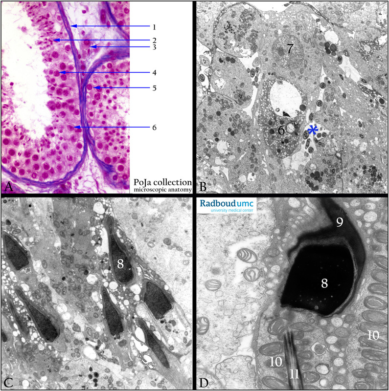
(A) Stain Azan, human. (B, C, D) Electron microscopy, gerbil.
(A-D): The survey (A) shows the seminiferous tubules with all cell types.
(C): Shows several maturing spermatozoa heads sticking into the Sertoli cell cytoplasm.
(D, 9): Acrosome cap covering the spermatozoon head. Helically arranged mitochondria (sheath) around the central axis of two
neighboring spermatozoa (D, 11) Axis of the middle piece forms the flagellum.
Keywords/Mesh: testis, seminiferous tubule, spermiogenesis, spermatozoon, acrosome, Sertoli cell, histology, electron microscopy, POJA collection
Title: Spermiogenesis in testes
Description:
(A) Stain Azan, human. (B, C, D) Electron microscopy, gerbil.
(A-D): The survey (A) shows the seminiferous tubules with all cell types.
- (1) Lamina propria limitans consisting of some myofibroblast cells.
- (2) Spermatids embedded in a Sertoli cell.
- (3) Interstitial Leydig cells with Reinke crystals.
- (4) Spermatocytes.
- (5) Spermatogonium.
- (6) Sertoli cell with a pale nucleus but a conspicuous nucleolus.
- (B, 6) Sertoli cell.
- (B, 7) Pre-Golgi phase spermatids.
(C): Shows several maturing spermatozoa heads sticking into the Sertoli cell cytoplasm.
(D, 9): Acrosome cap covering the spermatozoon head. Helically arranged mitochondria (sheath) around the central axis of two
neighboring spermatozoa (D, 11) Axis of the middle piece forms the flagellum.
Keywords/Mesh: testis, seminiferous tubule, spermiogenesis, spermatozoon, acrosome, Sertoli cell, histology, electron microscopy, POJA collection

