4.3.1 POJA-L-4201
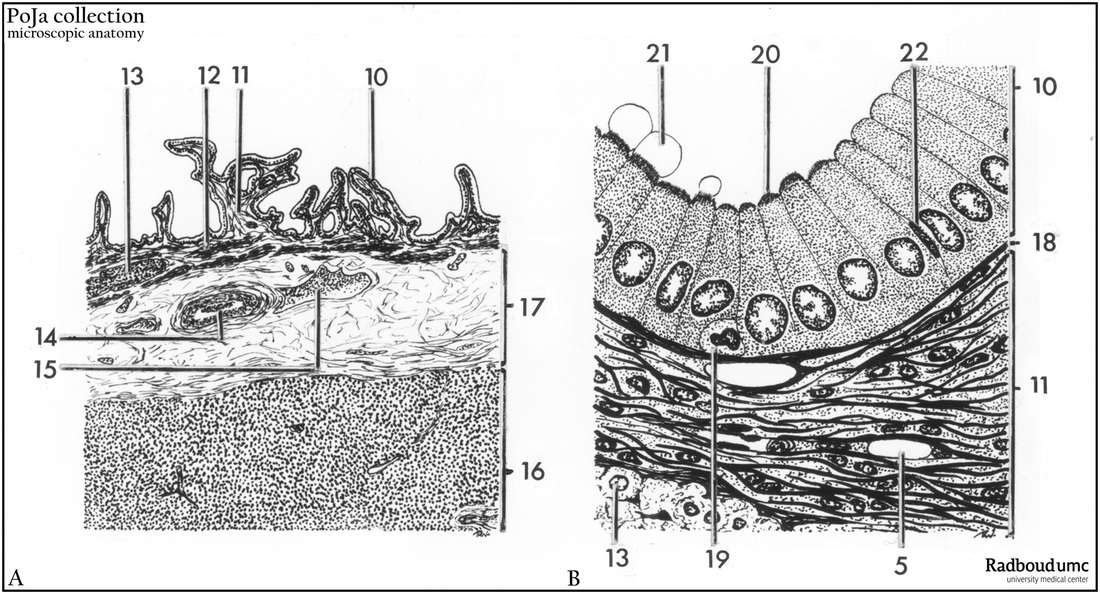
Title: Scheme gallbladder, Vesica fellea (vesica biliaris) (human)
Description: (A, B) Schematic survey and detail of light microscopic structure of gallbladder.
The gall bladder stores and concentrates bile produced in the liver and expels the bile in the duodenum via a common bile duct (ductus choledochus) from the liver (hepatic duct) and the gall bladder (cystic duct).
The mucosa of the gallbladder shows characteristic (branched) folds (plicae) (A, 10+11) with wide crypts in between. Deeper crypts are sometimes present, penetrating into the muscular layer and are known as diverticula or Rokitansky-Aschoff crypts. When contaminated with bacteria chronic inflammation might occur.
Simple columnar epithelium (A, B, 10) with short microvilli (20) or brush border delineate the lumen. Their cells are connected to each other by desmosomes and nexus structures. The apical side often shows bulging cytoplasmic protrusions or blebs (21, artifacts).
Infiltrated lymphocytes (19, diapedesis) can be observed at the basal side of the epithelium.
Below the basal membrane (18) a fine type, loosely built lamina propria (11) is found containing fibroblasts and free infiltrating lymphoid cells. The tunica fibromuscularis consists of a network of connective tissue fibers and helical smooth muscle fibers, cut transversely (13) or longitudinally (12).
The mentioned layers are surrounded by a loose connective tissue layer and a serosa or (17) tunica adventitia.
(14) Artery. (15) Veins with blood cells.
(16) Liver parenchyma. (22) Degenerating cell.
The irregular folding of the epithelium, the absence of goblet cells and the missing of a true muscularis mucosae makes it easy to discern a gallbladder histologically from a gut section. Cholecystokinin derived from entero-endocrine gut cells and released upon passage of fat, induces the contraction of the gallbladder. The bile, produced in the liver, is stored in the gallbladder and contains water and bile salts used to emulsify fat in the digestive tract.
Keywords/Mesh: gallbladder, bile duct, scheme, diverticulum, Rokitansky-Aschoff, histology, POJA collection

